XB-IMG-153102
Xenbase Image ID: 153102
|
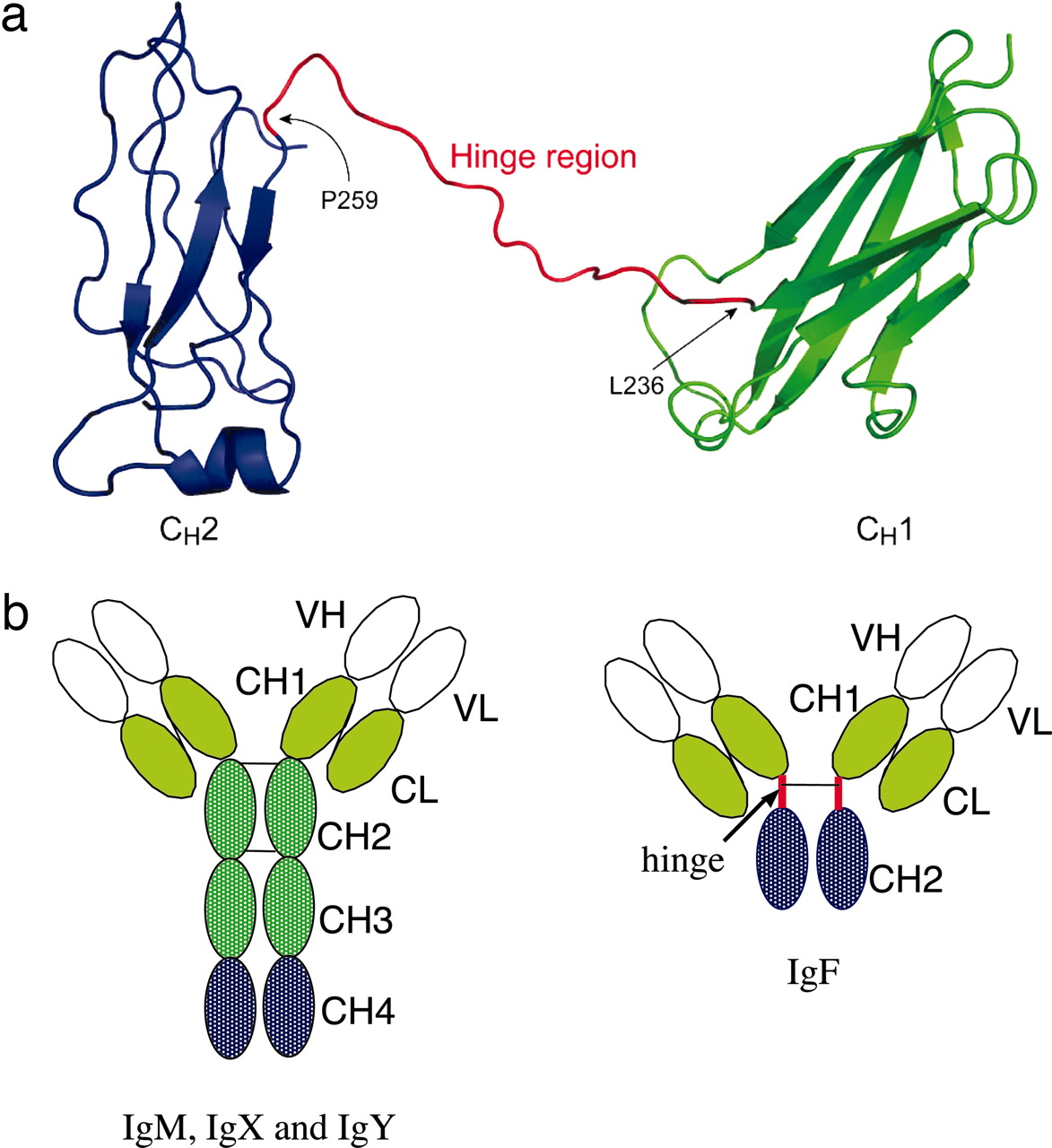
Fig. 4.
Structure of Igs in X. tropicalis. (a) A ribbon representation of the predicted structural model of the X. tropicalis IgF heavy chain. The CH1 and CH2 domains are colored green and blue, respectively. The putative hinge region between the two domains is colored red. Note that the hinge between CH1 and CH2 contains a gap (Ser-248 to Gly-252), which is due to the absence of corresponding residues in the template structure. The figure was prepared with PyMOL software. (b) Domain structure of IgF as compared with IgM, IgX, and IgY. There is only one cysteine in the C terminus of the CH2 domain of IgM for potential inter-heavy-chain disulfide bonding. CH, heavy-chain constant region domain; CL, light-chain constant region domain; VH, heavy-chain variable region; VL, light-chain variable region. Image published in: Zhao Y et al. (2006) Copyright © 2006. Image reproduced with permission of the publisher and the copyright holder. This is an Open Access article distributed under the terms of the Creative Commons Attribution License. Larger Image Printer Friendly View |