Click here to close Hello! We notice that you are using Internet Explorer, which is not supported by Xenbase and may cause the site to display incorrectly. We suggest using a current version of Chrome, FireFox, or Safari.
XB-IMG-153359

Xenbase Image ID: 153359


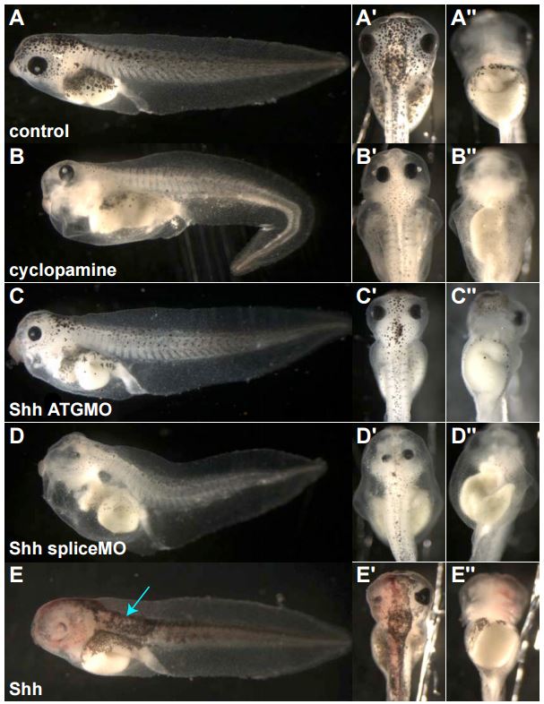
Fig. S3. Loss of Shh signaling and Hh signaling cause similar developmental defects. (A) Control embryos at stage 42, lateral (A), dorsal, (A') and ventral (A") views. (B) Cylopamine treatment blocks all Hh signaling, resulting in reduced melanocytes, edema, and kinked tail (B), closely spaced eyes (B’), and gut-looping defects (B"). Injection of MOs designed to the translation start site (“Shh ATGMO,” C–C") and splice donor site (“Shh spliceMO,” D–D") show similar phenotypes. (E) Injection of Shh mRNA (pink, lacZ staining) results in excess melanocytes (D, arrow), ventralization of the eye (E, E'), and gut looping defects (E").

Image published in: Peyrot SM et al. (2011)

Copyright © 2011. Image reproduced with permission of the Publisher, Elsevier B. V.

Experiment + Assay Source Phenotypes and Disease
Xla Wt + Cyclopamine + NF42 (morphology) fig.S3. b
Anatomical Phenotype
abnormal bending of tail
abnormally decreased number of melanophore
edematous ventral trunk
Xla Wt + Cyclopamine + NF42 (morphology) fig.S3. b^1
Anatomical Phenotype
abnormally localised eye
decreased size of the intercanthal region
Xla Wt + Cyclopamine + NF42 (morphology) fig.S3. b^1^1
Anatomical Phenotype
abnormal intestine morphology
Xla Wt + shh MO + NF42 (morphology) fig.S3.c
Anatomical Phenotype
abnormal bending of tail
abnormally decreased number of melanophore
edematous ventral trunk
Xla Wt + shh MO + NF42 (morphology) fig.S3.c'
Anatomical Phenotype
abnormal intestine morphology
Xla Wt + shh MO + NF42 (morphology) fig.S3.c^1
Anatomical Phenotype
abnormally localised eye
decreased size of the intercanthal region
Xla Wt + shh MO + NF42 (morphology) fig.S3.d
Anatomical Phenotype
abnormal bending of tail
abnormally decreased number of melanophore
edematous ventral trunk
Xla Wt + shh MO + NF42 (morphology) fig.S3.d'
Anatomical Phenotype
abnormal intestine morphology
Xla Wt + shh MO + NF42 (morphology) fig.S3.d^1
Anatomical Phenotype
abnormally localised eye
decreased size of the intercanthal region
Xla Wt + shh + NF42 (morphology) fig.S3.e
Anatomical Phenotype
abnormally increased number of melanophore
Xla Wt + shh + NF42 (morphology) fig.S3.e'
Anatomical Phenotype
abnormal development of alimentary system
abnormal intestine morphology
Xla Wt + shh + NF42 (morphology) fig.S3.e^1
Anatomical Phenotype
abnormally wholly ventralized eye

Larger Image
Printer Friendly View

Return to previous page