XB-IMG-153901
Xenbase Image ID: 153901
|
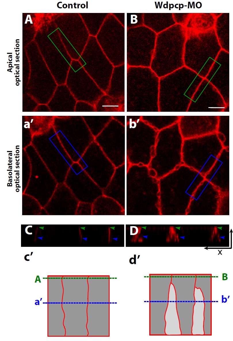
Fig. 2. Wdpcp knockdown caused severe membrane blebbing exclusively at the
basolateral membrane. (A) The apical plasma membranes in control Xenopus
epidermal epithelium, showing tight cell–cell contacts. (a0) The basolateral membranes
of cells shown in panel (A) are also remain stably connected (B). The apical
membrane of Wdpcp morphant tissue was indistinguishable from the control apical
membrane. (b0) Wdpcp morphants displayed severe blebbing of the basolateral
membranes. (C) X–Z projection of control epithelium. Green arrowheads indicate
apical junctions. Blue arrowheads indicate basolateral membrane. (D) X–Z projection
of Wdpcp morphant epithelium. Blue arrowheads indicate blebbing basolateral
membrane while the apical junction is quite stable (green arrowheads). The scale
bars are 10 lm. (c0 and d0) The drawing indicating the optical sectioning points for
confocal imaging of (A and B) (Green lines) or (a0 and b0) (Blue lines). Image published in: Park TJ et al. (2015) Copyright © 2015. This image is reproduced with permission of the publisher and the copyright holder. This is an Open Access article distributed under the terms of the Creative Commons Attribution License. Larger Image Printer Friendly View |