XB-IMG-154585
Xenbase Image ID: 154585
|
Figure 5
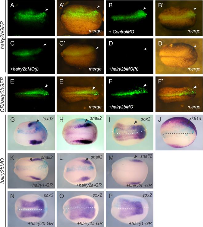
hairy2b is required for early neural crest specification in Xenopus embryos. A–P: Dorsal views. A–F: In vitro and in vivo efficiency of hairy2b antisense morpholino oligonucleotide (hairy2bMO). Embryos under a fluorescence stereo microscope; anterior side is on the right. White arrowheads indicate the injected side. A′,B′,D′,E′,F′: Fluorescence and clear field images of each embryo are shown in merged images. A–A′: Embryo injected with mRNA encoding hairy2bGFP (1 ng/embryo) showing GFP fluorescence on the treated side. B–B′: Embryo injected with hairy2bGFP mRNA (1 ng/embryo) and control antisense morpholino oligonucleotide (ControlMO, 20 ng/embryo). C–C′,D–D′: Embryos injected with hairy2bGFP mRNA (1 ng/embryo) and hairy2bMO (C–C′, low dose (l), 5 ng/embryo; D and D′, high dose (h), 10 ng/embryo). No embryo shows GFP fluorescence at a high dose of hairy2bMO. E,F: Embryo injected with mRNA encoding CRhairy2bGFP (E–E′, 1 ng/embryo) alone or co-injected with hairy2bMO (F–F′, high dose (h), 10 ng/embryo) showing GFP fluorescence on the treated side. G–P: Analysis of hairy2bMO effects on neural crest early specification. Dorsal views, anterior side is on the right. Black arrowheads indicate the injected side. G,H: hairy2bMO-injected embryos show inhibition of foxd3 and snail2 neural crest markers, respectively. I,J: The expression of the neural plate marker sox2 and the epidermal marker xk81a are expanded on the hairy2bMO-treated side. K,P: Co-injection of hairy2bMO and hairy1-GR or hairy2a-GR or hairy2b-GR mRNA rescues, snail2 (K–M) and sox2 (N–P) expression. The hairy genes rescue the expression of foxd3 in the neural crest (K–M) and the expression of sox2 in the neural plate (N–P). Image published in: Vega-López GA et al. (2015) Copyright © 2015. Image reproduced with permission of the Publisher, John Wiley & Sons.
Image source: Published
Larger Image Printer Friendly View |