XB-IMG-155162
Xenbase Image ID: 155162
|
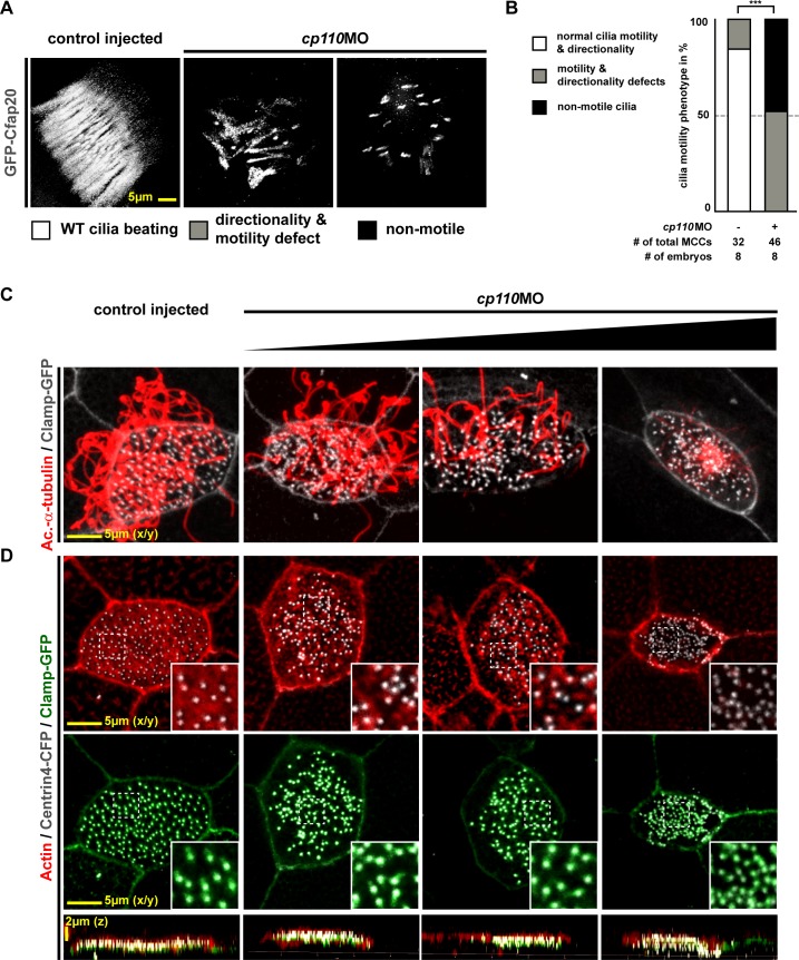
Figure 1. Cp110 is required for basal body function in MCC ciliogenesis.(A) cp110 knockdown causes impaired extracellular fluid flow. Control (uninjected controls and control MO injected specimens) and cp110MO-injected embryos were analyzed for extracellular fluid flow (10 s projections are shown). (B) Velocities were quantified by particle tracking (Related to Video 1). ***p<0.001; ns, p>0.05 from Wilcoxon two-sample test. (C) Alignment of basal bodies is disrupted in cp110 morphant MCCs. Centrin4-RFP (basal bodies, red), Clamp-GFP (rootlets, green). Arrows in bottom panels show basal body directionality. n embryos/MCCs: control (9/27), cp110MO (10/30). (D) Knockdown of cp110 causes severe defects in MCC ciliogenesis which can be rescued by cp110 DNA co-injection, demonstrated by immunofluorescence for Acetylated-α-tubulin (cilia, Ac.-α-tub., red). Trgeted MCCs were identified by co-injection of centrin4-cfp. Non-targeted MCCs (asterisks) produced normal cilia. (Related to Figure 1—figure supplement 2A). (E) Loss of Cp110 disrupts basal body apical transport and F-actin formation. Basal bodies (Centrin4-CFP, white) and Actin (red) are shown in apical (top row) and lateral (bottom rows) views of individual MCCs. Top views and lateral projections show representative examples (boxes indicate phenotype: white = wt; gray = mild docking defect; black = severe docking defect). (Related to Figure 1—figure supplement 2B,C). See also:DOI:
http://dx.doi.org/10.7554/eLife.17557.002Figure 1—figure supplement 1. Cp110 is required for basal body function in MCC ciliogenesis.(A) Cp110-deficient MCC cilia fail to beat directionally. gfp-cfap20 injected embryos were used to visualize ciliary beating (10 s projections are shown). (Related to Video 2–3). (B) Quantification of cilia motility data. ***p<0.001 from χ²-test. (C–D) cp110MO dose-dependent phenotypes of MCC ciliation, basal bodies and apical Actin. cp110MO doses used: 0pmol (control injected; first row), 3pmol (second row), 5pmol (third row), and 7pmol (fourth row). (C) Cilia were visualized by immunofluorescence for Acetylated-α-tubulin (cilia, Ac.-α-tub., red), rootlets were visualized by Clamp-GFP (white). n embryos/MCCs: control (6/19), cp110MO 3pmol (6/22), 5pmol (6/22), 7pmol (6/21). (D) Actin (red), basal bodies (Centrin-CFP, white), rootlets (Clamp-GFP, green). n embryos/MCCs: control (6/22), cp110MO 3pmol (6/17), 5pmol (6/22), 7pmol (6/29).DOI:
http://dx.doi.org/10.7554/eLife.17557.003 Image published in: Walentek P et al. (2016) © 2016, Walentek et al. This image is reproduced with permission of the journal and the copyright holder. This is an open-access article distributed under the terms of the Creative Commons Attribution license Larger Image Printer Friendly View |