XB-IMG-155211
Xenbase Image ID: 155211
|
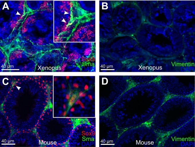
Fig. 4. Immunohistochemistry of agarose embedded testicular sections from X. tropicalis and mouse adult males. (A,C) Double staining with Sox9 (red) and Sma (green) antibodies. White arrowheads indicate potential common precursor cells for Sertoli and PTM cell lineages expressing both antigens in X. tropicalis (A) and mouse (C) samples. Insets show a detailed view of structures marked with white arrowheads in underlying figures. (B,D) Staining with vimentin (green) antibody on X. tropicalis (B) and mouse (D) samples. Nuclei were counterstained with DAPI (blue). Scale bars: 40â
μm. Image published in: Tlapakova T et al. (2016) © 2016. Creative Commons Attribution license
Image source: Published Larger Image Printer Friendly View |