XB-IMG-155603
Xenbase Image ID: 155603
|
Figure 1. Phosphorylation of Bub1 S459 is critical for spindle checkpoint activation.(A) Domains and motifs of Bub1 and sequence alignment of its conserved motif (CM). TPR, tetratricopeptide repeat; GLEBS, Gle2-binding sequence; Phe, phenylalanine-containing motif, also known as ‘ABBA’ motif; KEN, lysine-glutamate-asparagine motif. Hs, Homo sapiens; Mm, Mus musculus; Gg, Gallus gallus; Xl, Xenopus laevis; Dr, Danio rerio; Sp, Schizosaccharomyces pombe; Sc, Saccharomyces cerevisiae. The boxed regions in scBub1 and hsBub1 were synthesized as phospho-peptides and used in this study. (B) Recombinant Bub1ΔK–Bub3 complex was incubated with Cdk1–Cyclin B1 (Cdk1) or Mps1 in the presence of ATP. The kinase reactions were blotted with indicated antibodies. (C) HeLa cells stably expressing Myc-Bub1ΔK wild-type (WT) or S459A mutant were treated with nocodazole and MG132 in the presence or absence of reversine. Myc-Bub1 was immunoprecipitated and blotted with indicated antibodies. Endo., endogenous. (D) Flow cytometry of HeLa Tet-On cells stably expressing indicated siRNA-resistant Myc-Bub1 transgenes that were transfected with siBub1-d and treated with taxol. Mitotic indices were calculated as percentages of MPM2+ 4N cells in flow cytometry, and then plotted. FL, full-length. △K, mutant with the kinase domain truncated. Error bars, s.d. (n = 4 independent experiments). ****p<0.0001; Student’s t-test. (E) Representative flow cytometry plots of cells in (D). (F) HeLa Tet-On cells stably expressing indicated GFP-Bub1 transgenes were transfected with siBub1-d, treated with taxol, and imaged with time lapse microscopy. Cumulative percentages of cells remaining in mitosis were plotted against mitotic duration. Data from three independent experiments were combined. n (FL) = 161; n (△K)=173; n (△K S459A)=169. ****p<0.0001; Log-rank test. (G) Mitotic durations of cells stably expressing GFP-Bub1ΔK WT or S459A that were depleted of endogenous Bub1 and not treated with microtubule poisons. Data from three independent experiments were combined. Each dot represents one cell. n (WT) = 170; n (S459A)=157. ****p<0.0001; Student’s t-test.DOI:
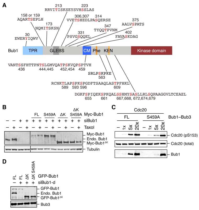
http://dx.doi.org/10.7554/eLife.22513.002Figure 1—figure supplement 1. Identification of mitotic phosphorylation sites in human Bub1 and characterization of the Bub1 S459A mutant.(A) Domain structure of human Bub1 and a summary of all 30 phosphorylation sites identified by mass spectrometry. Phosphorylated residues are denoted in red. (B) Lysates of cells in Figure 1D were blotted with anti-Bub1 and anti-Tubulin antibodies. (C) Recombinant Cdc20 was incubated with varying doses of recombinant Bub1–Bub3 full-length (FL) or S459A in the presence of ATP. The kinase reactions were resolved on SDS-PAGE and blotted with the indicated antibodies. (D) Lysates of cells in Figure 1F and G were blotted with anti-Bub1 and anti-Bub3 antibodies.DOI:
http://dx.doi.org/10.7554/eLife.22513.003 Image published in: Ji Z et al. (2017) © 2017, Ji et al. This image is reproduced with permission of the journal and the copyright holder. This is an open-access article distributed under the terms of the Creative Commons Attribution license Larger Image Printer Friendly View |