XB-IMG-156715
Xenbase Image ID: 156715
|
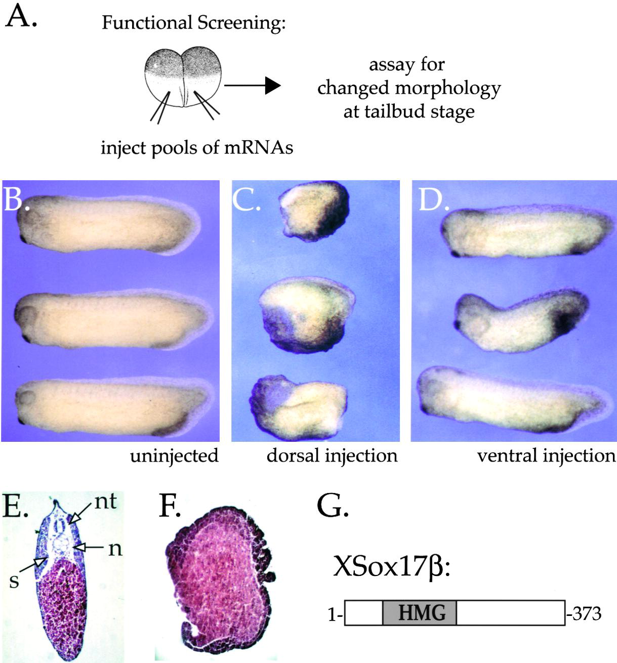
Figure 1.
Isolation of XSox17β
(A) Functional screen to identify genes regulating axis formation in Xenopus. Synthetic mRNA was prepared from pools of a dorsal lip cDNA library, and 2–5 ng was injected into 2–4 cell stage Xenopus embryos. Embryos were assayed for changes in morphology at the tail bud stage. A pool with ventralizing activity was serially subdivided and retested until a single active sequence was isolated. (B) Uninjected control embryos. (C) Dorsal injection of the active RNA (1 ng) resulted in ventralized embryos, while (D) ventral injection (1 ng) of the same RNA resulted in relatively normal development. Histological section of an uninjected control embryo (E) shows typical dorsal structures such as neural tube (nt), notochord (n), and somites (s), which are absent in the ventralized embryos (F). (G) The active clone encodes the HMG box protein, XSox17β. Image published in: Zorn AM et al. (1999) Copyright © 1999. Image reproduced with permission of the Publisher, Elsevier B. V.
Larger Image Printer Friendly View |