XB-IMG-157588
Xenbase Image ID: 157588

|
Figure 2
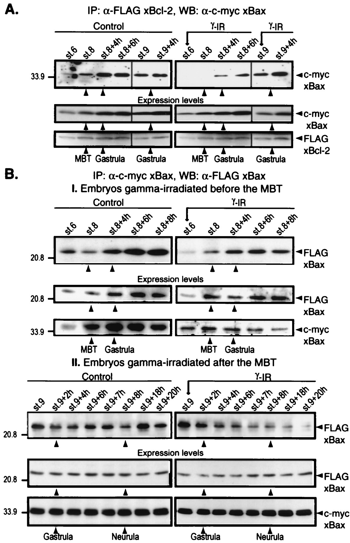
(A) Interaction between xBcl-2 and xBax changes at the MBT. Embryos were injected at the one-cell stage with a mixture of FLAG-tagged xBcl-2 and c-myc-tagged xBax mRNAs. The embryos were not irradiated (control) or were irradiated 3 h (stage 6) or 7 h (stage 9) after fertilization, collected at different times, and frozen. Samples equivalent to five embryos were immunoprecipitated with anti-FLAG M2-agarose beads, and the immunocomplexes were subjected to Western blot analysis with anti-c-myc antibody. (B) Analysis of xBax homodimerization. Embryos were injected at the one-cell stage with FLAG-tagged and c-myc-tagged xBax mRNAs, were not irradiated (control) or were irradiated at either stage 6 (I) or stage 9 (II), and were collected at different times. Samples equivalent to five embryos were immunoprecipitated with anti-c-myc-agarose beads, and the immunocomplexes were blotted with anti-FLAG M2 antibody (I, Top, and II, Top). Total FLAG-xBcl-2, FLAG-xBax, and c-myc-xBax expression levels were assessed at all stages by Western blotting with specific anti-tag antibodies (A and B, I and II, Middle and Bottom). Arrows on the right denote the FLAG-tagged or c-myc-tagged protein. Image published in: Finkielstein CV et al. (2001) Copyright © 2001. Image reproduced with permission of the Publisher and the copyright holder. This is an Open Access article distributed under the terms of the Creative Commons Attribution License. Larger Image Printer Friendly View |
