XB-IMG-157826
Xenbase Image ID: 157826
|
|||||||||||||||
|
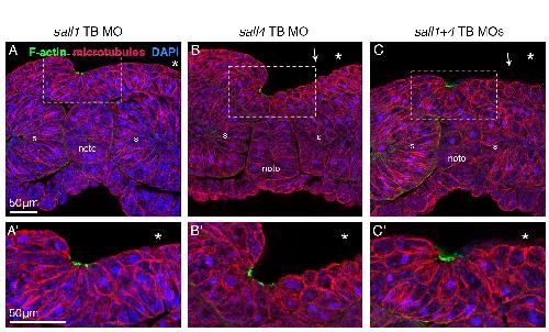
Supplementary Figure 7 â Apical actin accumulation is disrupted by translation-blocking morpholinos
Confocal microscopy after transverse sectioning and staining of morphants; dorsal is up. Sections were taken from the anterior third of embryos, where primary neurulation is most easily observed. F-actin (phalloidin, green), microtubules (anti-DM1α antibody, red), nuclei (DAPI, blue). Asterisks in images indicate injected side; white arrows indicate loss of actin accumulation. Aâ-Câ show higher magnification views of A-C. Scale bars = 50μm. s = somite, noto = notochord. Compare to Figure 3 F-Iâ, splice-blocking morpholinos; see Figure 3 J for quantification of phenotypes in all sall morphants.
Image published in: Exner CRT et al. (2017) Copyright © 2017. Image reproduced with permission of the Publisher, Elsevier B. V.
Image source: Published Larger Image Printer Friendly View |