XB-IMG-158376
Xenbase Image ID: 158376
|
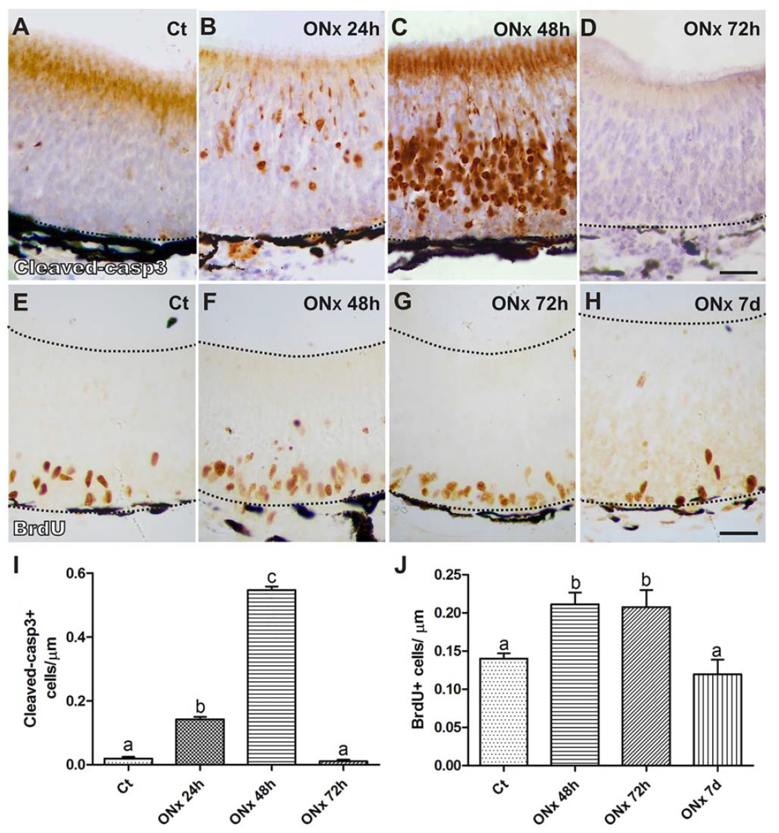
Figure 2 Axotomy triggers apoptosis in the OE and exacerbates proliferation of basal cells.
Apoptotic cells labeled with cleaved-casp3 (cleaved-casp31 cells) in the OE under normal
physiological conditions (A), and after 24 h (B), 48 h (C), and 72 h (D) post-axotomy; proliferating
cells labeled with BrdU (BrdU1 cells) in the OE under normal physiological conditions (E),
and after 48 h (F), 72 h (G) and 7 days (H) post-axotomy, scale bars: 20 mm; quantification of
cleaved-casp31 cells per mm of OE perimeter (I) and BrdU1 cells per mm of OE perimeter (J) in
control and axotomized animals after different recovery periods, different letters indicate statistically
significant differences between groups, n54 for each group, ANOVA and Tuckey test, P<0.05.
[Color figure can be viewed at wileyonlinelibrary.com] Image published in: Cervino AS et al. (2017) Copyright © 2017. Image reproduced with permission of the Publisher, John Wiley & Sons.
Image source: Published Larger Image Printer Friendly View |