XB-IMG-158744
Xenbase Image ID: 158744
|
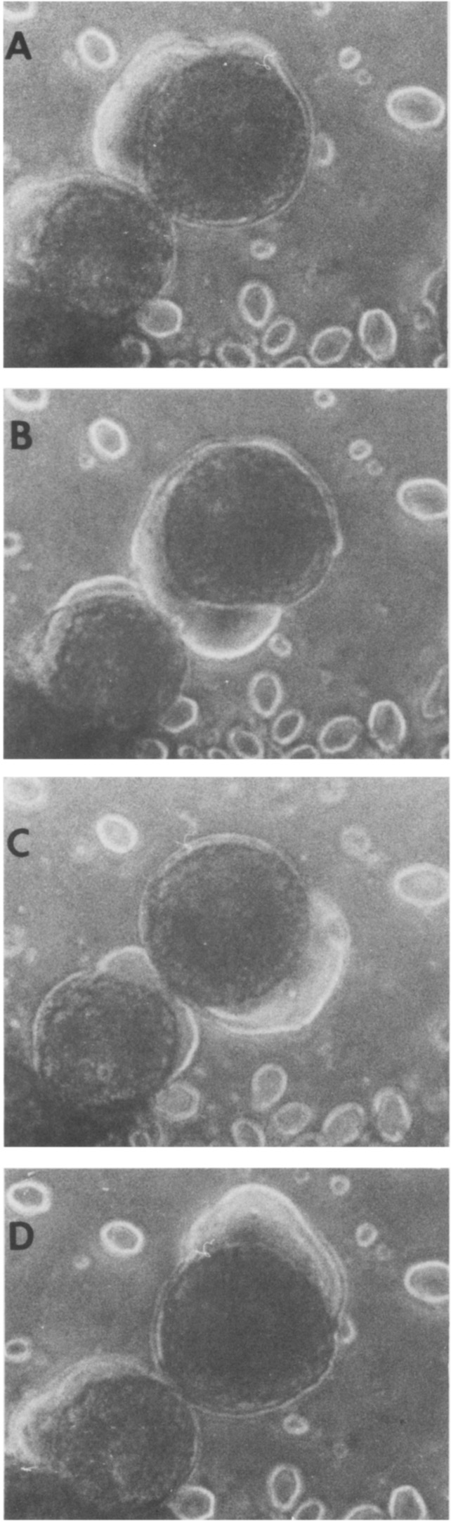
Figure 3. Demonstration of Motility Coincident with the MBT.
Embryos that had been growing in dissociation media were further disrupted by gentle shaking after cleavage 12. The cells were trans- ferred onto a microscope slide and photographed every 60 set at 160X. The small cytoplasmic bleb on the outer periphery of the cell will normally progress in a single direction around the cell for several revolutions before disappearing and then reappearing. (A) 0 min; (B) 1 min; (C) 2 min; (D) 3 min. At least 70%-80% of the cells defined as motile in Figure 2B exhibit this type of motility pattern. The remaining 20%-30% of motile cells undergo a much slower pseudopodal ex- tension and retraction motility. Final magnification 370X. Image published in: Newport J and Kirschner M (1982) Copyright © 1982. Image reproduced with permission of the Publisher, Elsevier B. V. Larger Image Printer Friendly View |