XB-IMG-159031
Xenbase Image ID: 159031
|
|
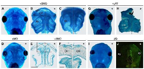
Figure S7. Vgll3 is required for the formation of neural crest derivatives. Embryos
were injected with 40 ng of v3MO (or cMO) or 1 ng of vgll3 mRNA (or gfp mRNA) and
fixed at stage 47 before alcian blue staining (A-I). (A, B, C, D) The formation of cranial
cartilage is impaired when vgll3 is knocked-down when compared to control MO
injected embryo [A compared to D (embryos injected with cMO show no alteration)].
This is more obvious on dissected embryos that revealed on the injected side a high
rate of cranial cartilage hypoplasia including severe loss of Meckel’s (MC), ceratohyal
(CH) and branchial cartilages (Br) (100%, n=15, Fig. 6B,C). (E, F) Transversal sections
of paraffin embedded tissues showed that the ceratohyal cartilage on the injected side
was shorter than on the uninjected control side (Fig. 6E) and displayed numerous
smaller chondrocytes (Fig. 6F, enlarged view of E indicated in the square line. Scale
bars represent 500 μm (E) and 130 μm (F). (G, H, I, J) Embryos injected with vgll3
mRNA showed an impaired cartilage development with the ceratohyal cartilage being
severely disorganized (100%, n=15, Fig. 6G, H). In control experiments, embryos
injected with gfp mRNA showed no change in cartilage head morphology (Fig. 6I, J).
ey, eye. Ventral views with anterior to the top (A, D, G, I) and dorsal views (J). (*)
injected side. Midline embryo is indicated by a dotted line. Image published in: Simon E et al. (2017) © 2017. This image is reproduced with permission of the journal and the copyright holder. This is an open-access article distributed under the terms of the Creative Commons Attribution license Larger Image Printer Friendly View |