XB-IMG-159175
Xenbase Image ID: 159175
|
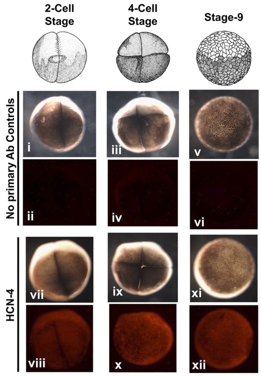
Figure 3: Xenopus laevis embryos express endogenous HCN4 channel during early
development.
Immunofluorescence analysis of whole Xenopus embryos for HCN4 channel
protein at indicated stages of development. (i-vi) no primary antibody controls (vii – xii)
HCN4 immunofluorescence. (i, iii, v, vii, ix, xi) Bright field images of immunofluorescent
embryos. (ii, iv, vi, viii, x, xii) Fluorescence images of immunofluorescence embryos.
Xenopus embryos at the indicated stage of development showed a prominent HCN4
channel protein (n=15). Image published in: Pai VP et al. (2017) © 2017. This image is reproduced with permission of the journal and the copyright holder. This is an open-access article distributed under the terms of the Creative Commons Attribution license
Image source: Published Larger Image Printer Friendly View |