XB-IMG-159359
Xenbase Image ID: 159359
|
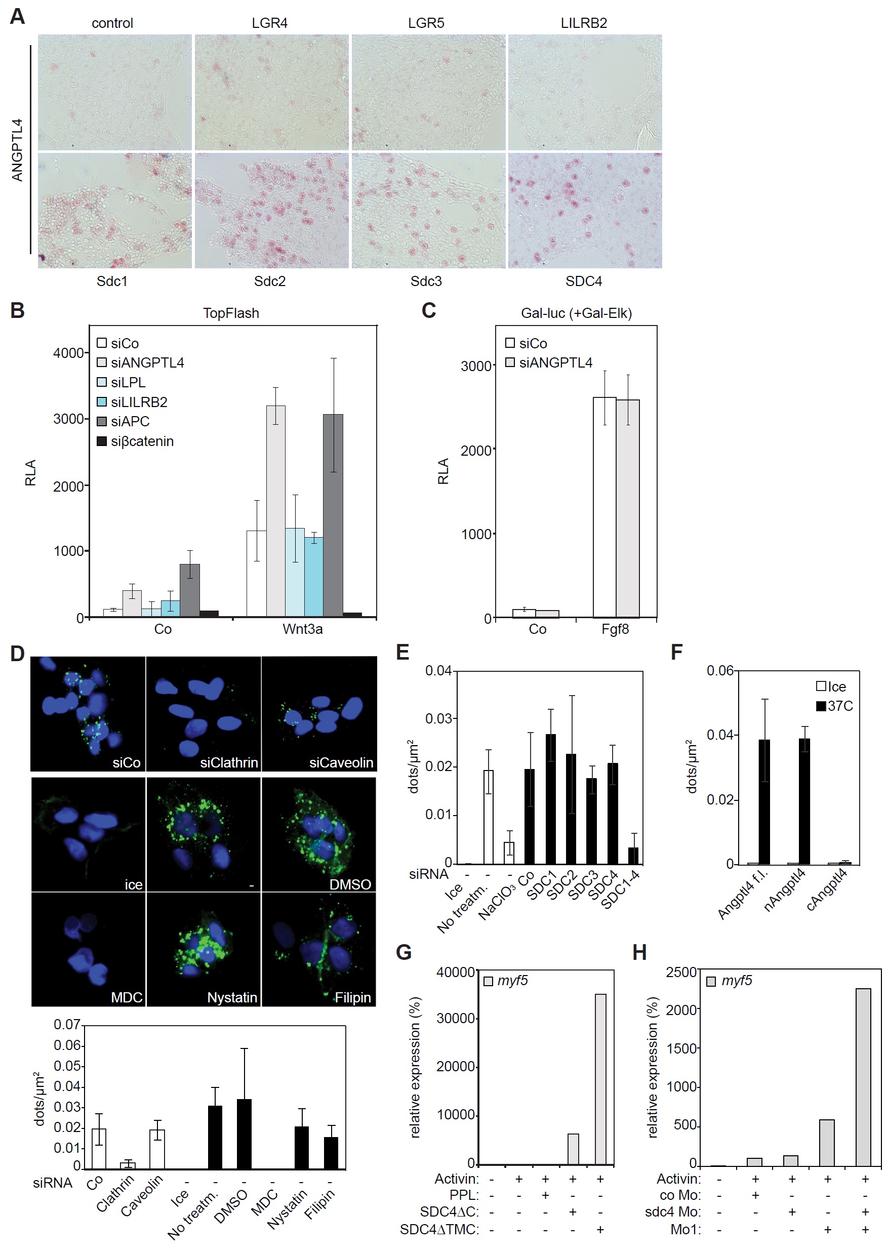
Figure S4, related to Figure 4. ANGPTL4 binds syndecans and is internalized by clathrin-dependent endocytosis.
(A) Cell surface binding assay in HEK293T cells. Cells were transfected with the indicated receptor constructs and incubated with conditioned medium containing ANGPTL4-AP (alkaline-phosphatase) fusion protein. Ligand binding was detected using a chromogenic AP substrate (red).
(B) Wnt luciferase reporter assay in H1703 cells in the presence of the indicated siRNAs.
(C) FGF reporter assay in HEK293T cells with binary Gal-Elk/Gal-luc reporter and
stimulated with recombinant mFgf8. Data (B and C) are displayed as mean ± SD, with 3
biological replicates. Co, control. RLA, relative luciferase activity.
(D) Internalization assay in HepG2 cells. Cells were treated with the indicated siRNAs or
endocytosis inhibitors, and incubated with HRP-tagged full-length ANGPTL4 (green). Protein
internalization was detected by tyramide signal amplification. MDC, monodansylcadaverine.
(E, F) Quantification of the indicated internalization assays shown in Fig. 4C (E) and Fig. 4D
(F). For internalization assays data are displayed as dots / μm² from multiple fields per
condition. Data are displayed as mean ± SD.
(G, H) qRT-PCR analyses of myf5 in animal cap explants from X. tropicalis embryos
microinjected as indicated. Note that the experiments belong to the data shown in Fig. 4G and
Fig. 4H respectively. Data show one representative of multiple independent experiments with
at least 3 biological replicates each. Image published in: Kirsch N et al. (2017) Copyright © 2017. Image reproduced with permission of the Publisher, Elsevier B. V. Larger Image Printer Friendly View |