XB-IMG-159755
Xenbase Image ID: 159755
|
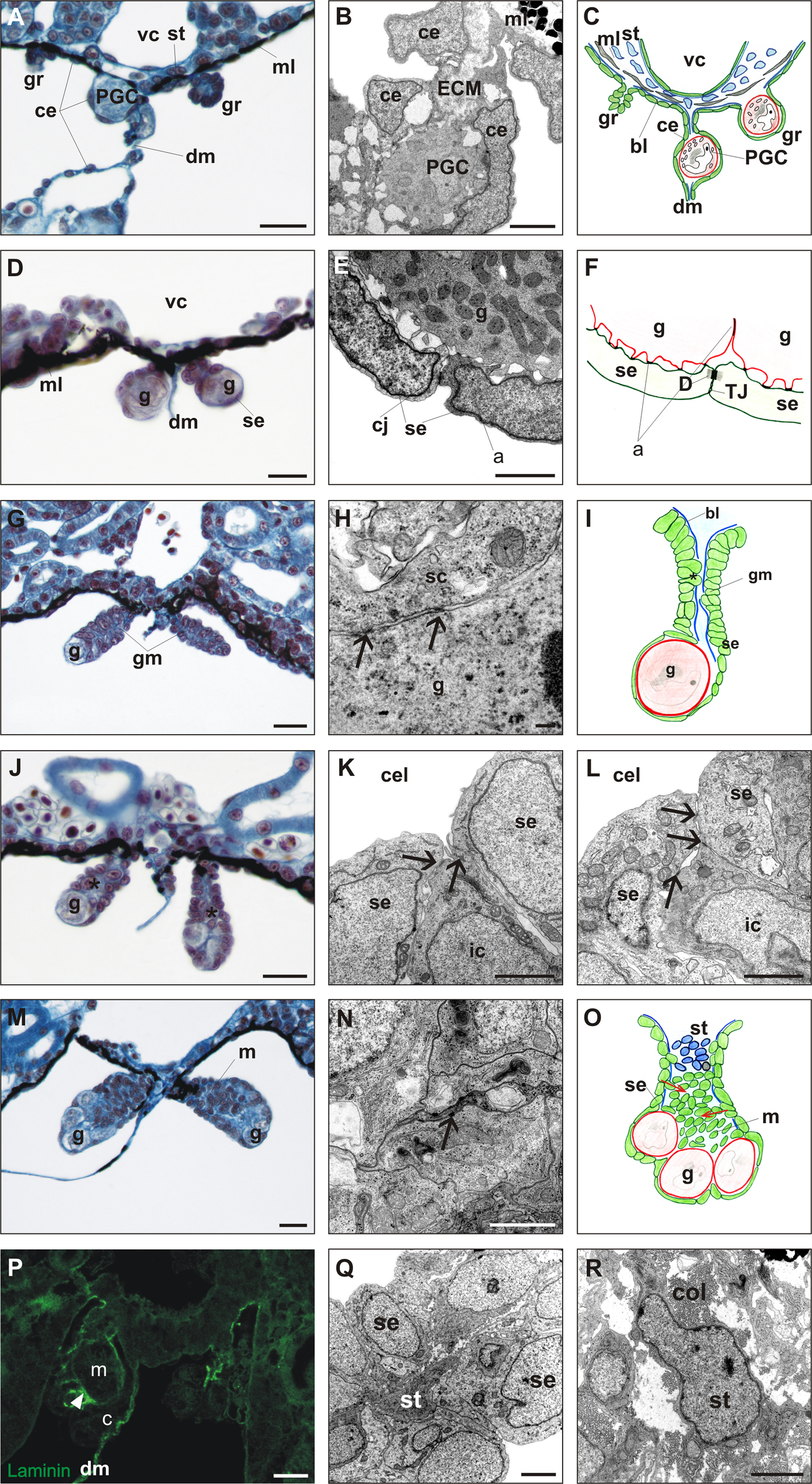
Fig. 1. The structure of undifferentiated gonad in Xenopus laevis. A. The beginning of the formation of genital ridges (gr) located on both sides of the dorsal mesentery (dm), just under vena cava (vc) at NF48. Primordial germ cells (PGC) during migration through dorsal mesentery towards the genital ridges. Stroma (st) and melanophores (mL) fill the space between vena cava and coelomic epithelium (ce). The genital ridges have a form of folded coelomic epithelium. B. Primordial germ cell (PGC) during migration through both sheets of dorsal mesentery formed by coelomic epithelium (ce). PGC adheres to the extracellular matrix (ECM) and coelomic epithelial cells without specialized cell junctions. C. Scheme of the genital ridge (left gr) before PGC settlement, the genital ridge with PGC (right gr), and PGC during migration through dorsal mesentery. Basal lamina (bl, blue line) disappears under coelomic epithelium in the sites of genital ridge formation. D. Undifferentiated gonads at NF49 are composed of flattened coelomic epithelial cells (gonadal surface epithelium, se) that cover germ cells (g). E, F. EM image and diagram of the germ cells adhering to the gonadal surface epithelium (se). Germ cells form microvilli adhering (a) to the somatic cells without specialized cell junctions. Cell junctions (cj) exist between adjacent somatic cells (tight junctions, TJ, and desmosome-like junctions, D). G. Undifferentiated gonads at NF50. Gonadal mesentery (gm) forms a proximal region of the gonad, and the germ cells are located in the distal region. First cells ingressing from the gonadal surface epithelium inwards the gonads are visible (asterisk). H. EM image of adjacent somatic (sc) and germ cell (g) at NF50. Both cells adhere to each other by electron dense points (arrows); basal lamina is absent. I. Diagram of undifferentiated gonads at NF50. The gonad has a form of monolayer epithelium enclosing germ cell (g) in the distal region; basal lamina (bl) is absent in the sites of contact with germ cell and ingressed somatic cells (asterisk). J. Undifferentiated gonads at NF51. Increase of the number of somatic and germ cells, and accumulation of somatic cells in the gonadal center (between both sheets of the gonad mesentery) (asterisk). K, L. EM images of ingression of cells from the gonadal surface epithelium toward the gonad center. Changes in the location of cell junctions are visible. K. Ingressing cell (ic) is the one of the gonadal surface epithelial cells (se); all cells are joined by cell junctions (arrows). L. Two surface epithelial cells (se) joined to each other above the ingressing cell (ic) which losses its connection with coelomic cavity (cel). Cell junctions (arrows). M. Undifferentiated gonads at NF52. In the center of the gonads the medulla forms a mass of somatic cells. Such undifferentiated gonads are composed of the cortex and medulla (m). All germ cells (g) are located exclusively at the periphery and are all joined to the gonadal surface epithelium. N. EM image of medulla cells forming a cluster of tightly packed cells joined by desmosome-like junctions. O. Diagram of undifferentiated gonads at NF52. Arrows indicate direction of cell ingression from the surface epithelium (se). P. Immunofluorescence of laminin reveals deposition of basal lamina (arrowhead) between the cortex (c) and medulla (m) in the undifferentiated gonads at NF52. Q. EM image of stromal cell (st) present between both sheets of surface epithelium in gonadal mesentery at NF52. R. EM image of stromal cell (st) in the undifferentiated gonad at NF53. The stromal cell is surrounded by abundant extracellular matrix containing mainly collagen fibers (col). Scale bars: A,D,G,J,M,P 20 µm; B,E 5 µm; K,L,N,Q,R 2 µm; H 200 nm.
Image published in: Piprek RP et al. (2017) Copyright © 2017. Image reproduced with permission of the Publisher, Elsevier B. V.
Image source: Published Larger Image Printer Friendly View |