XB-IMG-170409
Xenbase Image ID: 170409
|
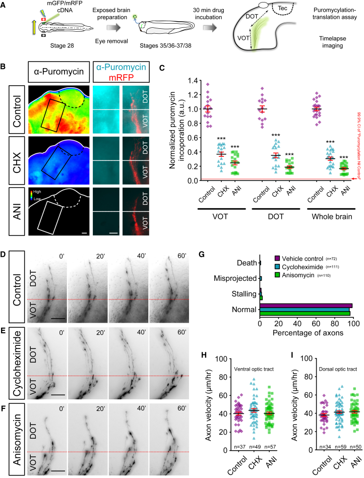
Figure 3. Axon Navigation in the Optic Tract Is Not Affected by Acute Inhibition of Translation(A) Live imaging experiment on somaless RGC axon navigation in the optic tract in vivo and translation assay on whole brains. Electroporated eye was removed to eliminate somatic contribution.(B) Anti-puromycin immunolabeling of whole-mount brains, shown as fluorescent intensity heatmaps, illustrates the incorporation of puromycin after 10 min, as readout of translation. Cycloheximide (CHX) and anisomycin (ANI) treatments greatly reduce puromycin immunolabeling.(C) The incorporation of puromycin was reduced in the ventral optic tract (VOT) (F3,67 = 204.6, p < 0.0001), dorsal optic tract (DOT) (F3,61 = 213.4, p < 0.0001), and whole brain (F3,80 = 501.9, p < 0.0001) after CHX and ANI treatments.(D–F) Axon navigation through the VOT and DOT in control (D) and after incubation in translation inhibitors CHX (E) and ANI (F).(G) Axon behaviors were unaffected in axons after CHX or ANI incubation (death: p = 0.44; misprojected: p = 0.19; stalling: p = 0.80; normal: p = 0.47, chi-square test).(H and I) Axon elongation velocities were unaffected by CHX or ANI incubation (H, VOT: F2,140 = 1.3, p = 0.29; I, DOT: F2,140 = 1.3, p = 0.27).Error bars represent SEM versus Control ∗∗∗p < 0.001 (one-way ANOVA with Tukey multiple comparison’s test for C, H, and I). Scale bars, 50 μm. Image published in: Wong HH et al. (2017) © 2017 The Authors. This image is reproduced with permission of the journal and the copyright holder. This is an open-access article distributed under the terms of the Creative Commons Attribution license Larger Image Printer Friendly View |