XB-IMG-170440
Xenbase Image ID: 170440
|
|
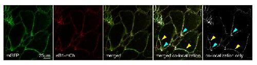
Figure 9—figure supplement 2. Co-localization of ephrinB1 (eB1-mCh) with membrane-label (mGFP) shows that ephrinB1 is dispersed over the entire membrane, but enriched at the trailing edge membrane (yellow arrows), particularly at the rear (blue arrows).Individual images are shown alongside the merged image. From left to right, the images represent mGFP only, eB1-mCh only, merged composite, a co-localisation rendering showing sites of co-localisation in white (white areas) superimposed on a composite image of mGFP and eB1-mCh, and a co-localisation-only panel, with the composite image removed for clarity. Image published in: Wen JW and Winklbauer R (2017) © 2017, Wen et al. This image is reproduced with permission of the journal and the copyright holder. This is an open-access article distributed under the terms of the Creative Commons Attribution license Larger Image Printer Friendly View |