XB-IMG-170727
Xenbase Image ID: 170727
|
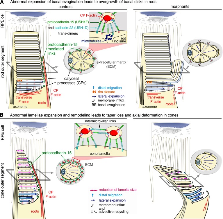
Figure 9. Model of the structural defects observed in the photoreceptors of morphant larvae. (A and B) In rod (A) and cone (B) photoreceptor cells, the basal evaginations (BEs) are formed with their leading edge exposed to the extracellular space. Calyceal processes (CPs) filled by F-actin (red) surround the base of the outer segment (OS) in control rods and cones (A and B, left). Links formed by trans-interactions between protocadherin-15 and cadherin-23 likely couple CPs to the OS membrane and to other CPs in the cones. The formation of trans-homotypic links, made up of either protocadherin-15 (Indzhykulian et al., 2013) or cadherin-23 cannot be excluded (A and B). (A, top left) Rod BEs form a continuous membrane system, topologically equivalent to cone lamellae. Lateral expansion of the BEs is tightly coupled to disk formation and distal displacement. Rim closure creates a discontinuity between the disk and the plasma membrane, precluding membrane redistribution in the plasmalemma. (top right) Uncontrolled lateral expansion of the nascent disks might lead to aberrant outgrowths, as shown in the pcdh15 morphant rods. In the morphant rods, the internal cytoskeleton of microtubules along the incisures may sustain the persisting CPs, although the F-actin cytoskeleton within the processes is markedly decreased. (B) Unlike rods, cones have no incisures, and the CPs do not anchor to microtubules. (bottom left) Preservation the cone OS shape requires a balanced distribution of forces and flows. Membrane input into the membrane folds of the cone lamellae at the OS base is equilibrated by advective recycling in the plasmalemma and apical shedding of older lamellae. This results in the lateral expansion, distal displacement, and progressive resizing of lamellae by retinal pigment epithelium (RPE) cells. (bottom right) In pcdh15 morphant cones lack CPs; disturbance of the balance between these antagonistic forces causes taper loss and an abnormal axial curvature of the OS. Image published in: Schietroma C et al. (2017) © 2017 Schietroma et al. This image is reproduced with permission of the journal and the copyright holder. This is an open-access article distributed under the terms of the Creative Commons Attribution-NonCommercial-ShareAlike license Larger Image Printer Friendly View |