XB-IMG-172641
Xenbase Image ID: 172641
|
Fig. S1.
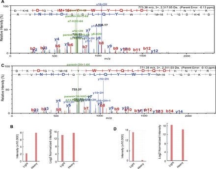
Mass spectrometry quantification of a RSF1 unique peptide in H2Aub-enriched and -depleted nucleosomes. (A) Spectrum of a RSF1 fragment (DKDGLmYWYQLDQDHNVR) in H2Aub-enriched and -depleted nucleosomes. In these experiments, H2Aub-enriched nucleosomes were labeled with heavy lysine and H2Aub-depleted nucleosomes were obtained from cells cultured in light medium. (B) Quantification of the labeled RSF1 fragment intensity from A. Both the intensity and log2-normalized intensity are shown. (C) Spectrum for RSF1 Fragment (DK+6DGLmYWYQLDQDHNVR) in H2Aub-enriched and -depleted nucleosomes. In these experiments, H2Aub-depleted nucleosomes were labeled with heavy lysine and H2Aub-enriched nucleosomes were obtained from cells cultured in light medium. (D) Quantification of the labeled RSF1 fragment intensity from C. Both the intensity and log2-normalized intensity are shown. Image published in: Jones AE et al. (2017) Copyright © 2017. Image reproduced with permission of the publisher and the copyright holder. This is an Open Access article distributed under the terms of the Creative Commons Attribution License. Larger Image Printer Friendly View |