XB-IMG-172926
Xenbase Image ID: 172926
|
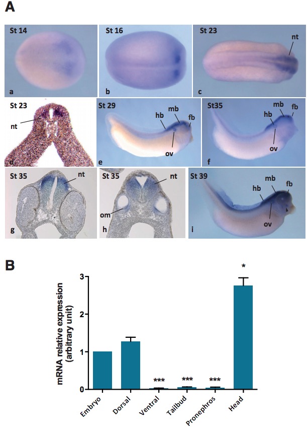
Fig. 5 (right). Spatio-temporal expression of pou3f2 during Xenopus laevis development. (A) Whole mount in situ hybridization for pou3f2 at
indicated stages of development. Neurula, dorsal view (a,b). Tailbud, dorsal (c) and lateral (e) view. Tadpole, lateral view (f,i). Transverse sections at the
level of the developing brain (d,g,h), and otic vesicle (h). Abbreviations: (fb) forebrain, (hb) hindbrain, (mb) midbrain, (nt) neural tube, (om) otic mesenchyme,
(ov) otic vesicle. (B) Expression of pou3f2 analyzed by real-time quantitative polymerase chain reaction (RT-qPCR) on dissected explants from
tailbud embryos (stage 25). Embryos were dissected either into ventral and dorsal halves or into tailbud, pronephros and head. *: P < 0,05; ***: P <
0,001, relative to the whole embryo. Image published in: Cosse-Etchepare C et al. (2018) Copyright © 2018. Image reproduced with permission of the Publisher, University of the Basque Country Press. Larger Image Printer Friendly View |