XB-IMG-173203
Xenbase Image ID: 173203

|
|
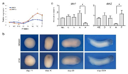
Figure 4. Developmental expression of dkk2. (a) Temporal expression of dkk2.L and dkk2.S by qRT-PCR. (b) By in situ hybridization, at all stages
examined (NF stage 11-33/34) dkk2 does not appear to be spatially restricted. Sense probe is shown as a control. (c) qRT-PCR analysis of dkk1 and dkk2 expression in dissected embryos at stage 15. WE; whole embryo; D, dorsal half; V, ventral half; A, anterior half; P, posterior half. The values were
normalized to Ef1a and presented as mean ± s.e.m. * p<0.05, Student’s t-test. Image published in: Devotta A et al. (2018) © 2018, Devotta et al. This image is reproduced with permission of the journal and the copyright holder. This is an open-access article distributed under the terms of the Creative Commons Attribution license Larger Image Printer Friendly View |
