XB-IMG-174703
Xenbase Image ID: 174703
|
|||||||||||||||||||||||||||||||||||
|
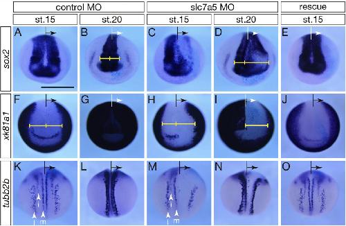
Fig. 7.Inhibition of slc7a5 led to the disorganization of neural- and non-neural patterning and primary neurogenesis.Neurula stage embryos (st.15 and st.20) were surveyed to analyze neural- and non-neural patterning in slc7a5-depleted embryos. Whole-mountin situhybridizationwas performed withsox2,xk81a1,tubb2b(N-tubulin) probes in embryos injected with control MO or slc7a5 MO. Black and white arrow represents the injected side.Yellow brackets indicate the width of the neural region in the embryo. (AâE)sox2expression. Anterior view. Broader expression domain was observed in slc7a5 MO-injected side. (FâJ)xk81a1expression. Anterior view.xk81a1-negative domain was observed in the anterior part of slc7a5 MO-injected side even after st.20. (KâO)tubb2bexpression. Dorsal view. Motoneuron in medial region and interneuron in more lateral region were eliminated in slc7a5 MO-injected embryo. (E, J, O)Coinjection ofslc7a5.Sandslc7a5.LmRNA with slc7a5 MO rescued these phenotypes.β-GalactosidaseRNA (1 ng) was used as a tracer. Scale bars: 1 mm. l: lateralneurons (sensory neurons or Rohon-Beard neurons), i: intermediate neurons (interneurons), m: medial neurons (motoneurons). (For interpretation of the referencesto color in thisfigure legend, the reader is referred to the web version of this article.) Image published in: Katada T and Sakurai H (2019) Copyright © 2019. Image reproduced with permission of the Publisher, Elsevier B. V.
Image source: Published Larger Image Printer Friendly View |