XB-IMG-1755
Xenbase Image ID: 1755
|
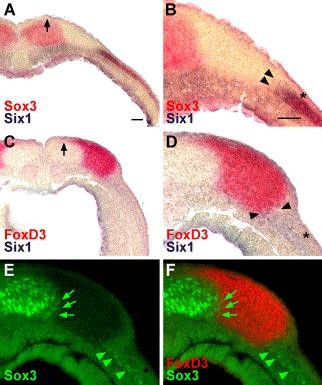
Fig. 6. Positioning of gene expression domains at the lateral neural plate border as revealed by double-staining procedures in transverse sections through the cranial neural folds of neural fold stage Xenopus embryos (stages 16–17). (A and B) Double in situ hybridization reveals a gap between the neural plate domain of Sox3 expression and the lateral crescent of Six1 expression. Arrow in A indicates tip of the neural folds. Arrowheads in B (representing higher magnification of A) indicate medial border of Six1 expression in the deep ectodermal layer (superficial ectodermal layer marked by asterisk). (C–F) Double in situ hybridization for FoxD3 (red) and Six1 (blue; medial border of expression marked by arrowheads in D) combined with Sox3 immunohistochemistry (E) reveals the position of the FoxD3-expressing neural crest relative to the Sox3-immunopositive neural plate and the lateral, placodal region that expresses Six1 and is immunoreactive for Sox3. Arrow in C indicates the tip of the neural folds. Arrowheads in D (representing higher magnification of C) indicate the medial border of Six1 expression in the deep ectodermal layer (superficial ectodermal layer marked by asterisk). Green arrows in E and F (showing a superposition of a fluorescent image of FoxD3 expression with E) indicate that Sox3-immunopositive nuclei of the lateral neural plate are located within the domain of FoxD3 expression. Green arrowheads in E and F indicate Sox3-immunopositive nuclei in the deep ectodermal layer of the lateral, placodal domain, which also expresses Six1 (compare with D), but does not show clear overlap with FoxD3 expression. Scale bar in A: 100 μm (for A and C). Scale bar in B: 100 μm (for B and D–F). Image published in: Schlosser G and Ahrens K (2004) Copyright © 2004. Image reproduced with permission of the Publisher, Elsevier B. V.
Image source: Published Larger Image Printer Friendly View |