Click here to close Hello! We notice that you are using Internet Explorer, which is not supported by Xenbase and may cause the site to display incorrectly. We suggest using a current version of Chrome, FireFox, or Safari.
XB-IMG-176578

Xenbase Image ID: 176578

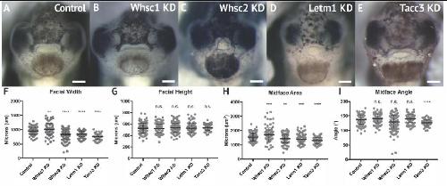
FIGURE 3. WHS related gene depletion affects craniofacial morphology. (A–E) Frontal views of 3dpf embryos (st. 40) following WHS gene single KD. (F–I) Measurements for facial width, height, midface area, and midface angle. A significant 6.54% increase in facial width and 11.43% increase in midface area were observed for Whsc1 KD. Whsc2 KD caused a 12.01% reduction in facial width and a 6.79% reduction in midface area. Letm1 KD caused a 10.33% decrease in facial width and a 8.49% decrease in midface area. Tacc3 KD caused a 21.27% decrease in facial width and a 16.33% decrease in midface area, and an 8.27% decrease in midface angle. Significance determined using a student’s unpaired t-test. (Embryos quantified: Control = 137, Whsc1 KD = 100, Whsc2 KD = 185, Letm1 KD = 115, Tacc3 KD = 79.) ∗∗∗∗P < 0.0001, ∗∗∗P < 0.001, ∗∗P < 0.01, n.s., not significant. Scalebar = 250 μm.

Image published in: Mills A et al. (2019)

Copyright © 2019 Mills, Bearce, Cella, Kim, Selig, Lee and Lowery. This image is reproduced with permission of the journal and the copyright holder. This is an open-access article distributed under the terms of the Creative Commons Attribution license

Experiment + Assay Source Phenotypes and Disease
Xla Wt + nsd2 MO + NF40 (whole-mount microscopy) Fig. 3B, FGHI
Anatomical Phenotype
abnormal craniofacial region morphology
increased size of the head
Disease
Wolf-Hirschhorn syndrome
Xla Wt + nelfa MO + NF40 (whole-mount microscopy) Fig. 3C, FGHI
Anatomical Phenotype
abnormal craniofacial region morphology
decreased size of the head
Disease
Wolf-Hirschhorn syndrome
Xla Wt + letm1 MO + NF40 (whole-mount microscopy) Fig. 3D, FGHI
Anatomical Phenotype
abnormal craniofacial region morphology
decreased size of the head
Disease
Wolf-Hirschhorn syndrome
Xla Wt + tacc3 MO + NF40 (whole-mount microscopy) Fig. 3E, FGHI
Anatomical Phenotype
abnormal craniofacial region morphology
decreased size of the head
Disease
Wolf-Hirschhorn syndrome

Larger Image
Printer Friendly View

Return to previous page