XB-IMG-176581
Xenbase Image ID: 176581
|
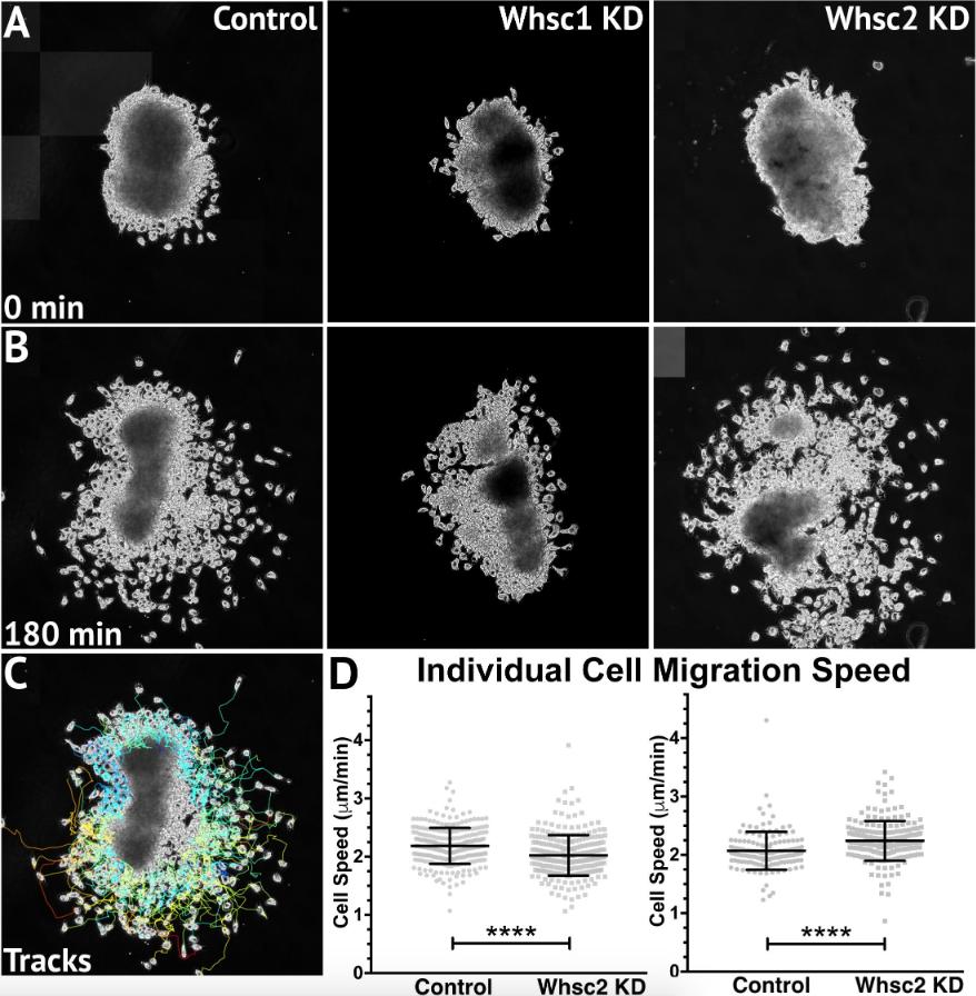
FIGURE 6. Whsc1 manipulation alters CNC migration speeds in vitro. Dissected CNC explants from control, Whsc1 KD, or Whsc2 KD embryos were plated on fibronectin-coated coverslips, allowed to adhere and begin migration, and imaged for 3 h using 20à phase microscopy. (A) Representative explants at initial timepoint (0 min). (B) Explants after 3 h migration time. (C) Representative tracks generated by FiJi Trackmate plug-in. (D) Mean track speeds of Whsc1 or Whsc2 KD explants compared to their controls. (Explants quantified: 3â4 explants from control and KD embryos were plated for each experiment, explants with neural or epithelial contaminant were excluded from analysis. Three separate experiments were performed for each depletion. Whsc1 controls: 272 cells, 9 explants. Whsc1 KD: 282 cells, 9 explants. Whsc2 controls: 151 cells, 12 explants. Whsc2 KD: 195 cells, 8 explants.) ââââP < 0.0001, n.s., not significant. Scalebar is 250μm. Image published in: Mills A et al. (2019) Copyright © 2019 Mills, Bearce, Cella, Kim, Selig, Lee and Lowery. Creative Commons Attribution license
Larger Image Printer Friendly View |