XB-IMG-176588
Xenbase Image ID: 176588
|
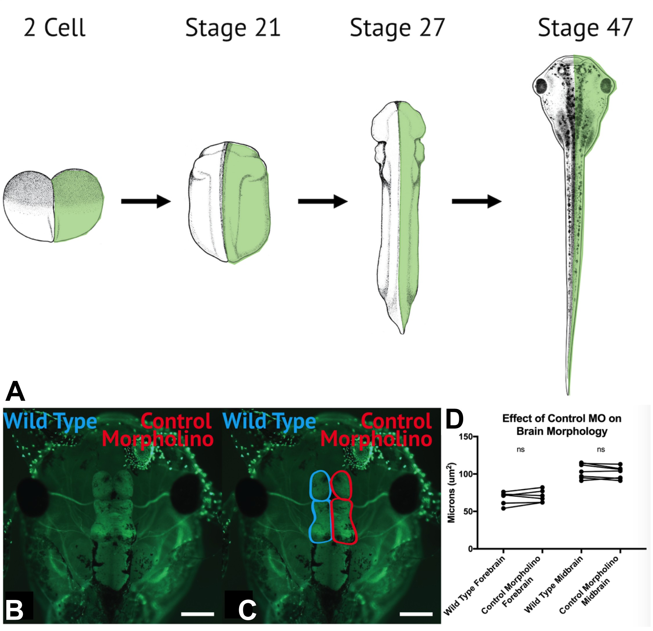
FIGURE S5 | Half embryo knockdown can be utilized for analysis of brain morphology and neural crest cell migration in vivo. (A) At the 2-cell stage, a single blastomere is injected with WHS-associated gene MOs and exogenous eGFP mRNA. After neurulation (approx. stage 21), embryos are sorted based on left or right eGFP fluorescence, to determine side of depletion. To examine neural crest cell size, migration and morphology embryos were fixed between stage 25â30, and in situ hybridization was performed using twist anti-sense probe. To characterize brain morphology, embryos were raised to st. 47, and fixed and labeled with alpha-tubulin antibody, a neuronal marker, and Alexa-488 secondary. (BâD) Control MO does not significantly impact brain size, compared to non-injected hemispheres (a paired internal control). Image published in: Mills A et al. (2019) Copyright © 2019 Mills, Bearce, Cella, Kim, Selig, Lee and Lowery. Creative Commons Attribution license Larger Image Printer Friendly View |