XB-IMG-177597
Xenbase Image ID: 177597
|
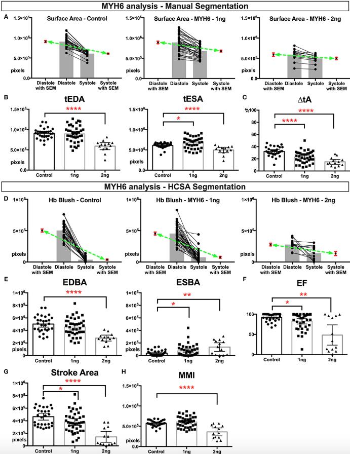
Figure 1. Physiological quantification of embryo heart function in MYH6 morphants. For this experiment 29 control, 39 morphants-−1 ng morpholino injected and 13 morphants-−2 ng morpholino injected analyzed. Measurements derived from manual segmentation include surface area at the end of diastole and systole and the change in ventricle area. (A) Before-after graph presented with extra columns pointing mean with SEM in red. Green line between means flattens as change in surface area diminishes with increasing MO dose. (B,C) As the MO dose is increased, the change in ventricle area is reduced, and the ventricle sizes are smaller. (D) Measurements derived from HCSA application include Hb blush at the end of diastole and systole and the change in blush area. Before-after graph presented with extra columns pointing mean with SEM in red. Green line between means flattens as change in blush area diminishes with increasing MO dose. (E,F) Following HCSA application, Hb blush is quantified at the end of diastole and systole. Ejection Fraction is derived from these measurments. Morphants demonstrated a dose dependent diminished ejection fraction and (G) stroke area. (H) Myocardial mass index estimates the amount of cardiac mass within the manually segmented heart. Myocardial mass index is affected in 2 ng morphants but remains within normal limits in 1 ng morphants. SEM, standard error of the mean; Hb Blush, Hemoglobin-containing pixels; tEDA, total end-diastolic area; tESA, total end-systolic area; ΔtA, change in total area; EDBA, end diastolic blood area; ESBA, end-systolic blood area; EF, ejection fraction; MMI, myocardiac mass index; HCSA, Hb contrast subtraction angiography. ****p < 0.0001, **p < 0.01, and *p < 0.05. Image published in: Deniz E et al. (2019) Copyright © 2019 Deniz, Jonas, Khokha and Choma. This image is reproduced with permission of the journal and the copyright holder. This is an open-access article distributed under the terms of the Creative Commons Attribution license Larger Image Printer Friendly View |