XB-IMG-1807
Xenbase Image ID: 1807
|
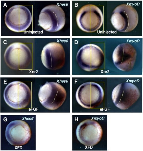
Fig. 1. Xhes6 is induced by mesoderm inducing signals and its expression is dependent on FGF-signaling. 5 pg of Xnr2 (C, D), 1 pg of eFGF (E, F) or 125 pg of XFD (G, H) mRNA was injected into a blastomere at 2-cell stage, along with β-galactosidase mRNA (red staining) and analyzed at gastrula stage for expression of Xhes6 (A, C, E and G) and XmyoD (B, D, F and H) by whole-mount in situ hybridisation. The side view of the area within the yellow box is shown on the right of each panel. Image published in: Murai K et al. (2007) Copyright © 2007. Image reproduced with permission of the Publisher, Elsevier B. V.
Image source: Published Larger Image Printer Friendly View |