XB-IMG-23258
Xenbase Image ID: 23258
|
|||||||||||||||||||||||||
|
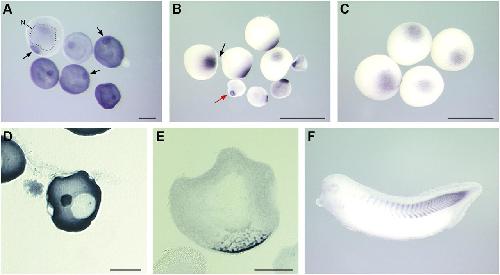
Fig. 2. XNIF encodes for a vegetally localized RNA in Xenopus oocytes. Whole-mount in-situ hybridization was carried out using albino oocytes of different stages. For a detailed analysis, sections of stained stage I and stage II oocytes were prepared. (A) In stage I oocytes, XNIF RNA is enriched in the mitochondrial cloud (examples marked by arrows). The cell nucleus is indicated by a dashed line in one oocyte. (B) In stage II oocytes, XNIF accumulates at the tip of the vegetal cortex (red arrow). In stage III-IV oocytes, XNIF RNA is associated with the vegetal cortex (black arrow). (C) Vegetal-cortical localization of XNIF in stage V/VI oocytes. (D) Section of whole-mount in-situ stained stage I oocyte. XNIF transcript is enriched in the mitochondrial cloud adjacent to the germinal vesicle. (E) Section of whole-mount in-situ stained stage II oocyte. XNIF is associated with reticular structures in a wedge-shaped region beneath the germinal vesicle. Scale bar:∼ 100 μm in (A,D,E) and 1 mm in (B,C). (F) XNIF expression in a stage 33 Xenopus albino embryo. Whole-mount in-situ hybridization staining with a XNIF specific antisense probe is shown. Image published in: Claussen M et al. (2004) Copyright © 2004. Image reproduced with permission of the publisher and the copyright holder. This is an Open Access article distributed under the terms of the Creative Commons Attribution License.
Image source: Published Larger Image Printer Friendly View |