XB-IMG-23717
Xenbase Image ID: 23717

|
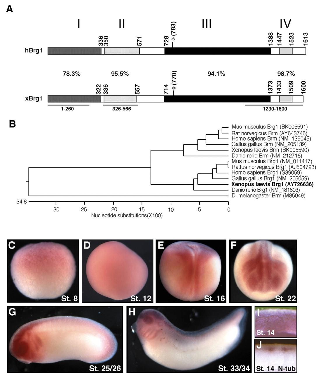
Fig. 1. Cloning and expression profile of xBrg1. (A) Structure of hBrg1 and xBrg1. Domains are labeled after (Khavari et al., 1993) with percent amino acid identity shown. Asterisks mark the ATP binding pocket targeted by mutagenesis to generate a dominant-negative form. Lines under xBrg1 indicate probes used for in situ hybridization. (B) Phylogenetic analysis of Brg1 and Brm orthologs. Units indicate the number of substitutions. Distance between any two sequences is the sum of horizontal branch length separating them. (C-I) Expression profile of xBrg1. (C) Stage 8 and (D) stage 12, side views (vegetal pole toward bottom). (E) Dorsal view, anterior towards the top (stage 16). (F) Anterior view (stage 22). (G) Stage 25/26 and (H) stage 33/34, lateral views. (IJ) Cross-sectional views of stage 14 embryos stained for xBrg1 (I) or N-tubulin (J). Image published in: Seo S et al. (2005) Copyright © 2005. Image reproduced with permission of the publisher and the copyright holder. This is an Open Access article distributed under the terms of the Creative Commons Attribution License.
Image source: Published Larger Image Printer Friendly View |
