XB-IMG-23828
Xenbase Image ID: 23828
|
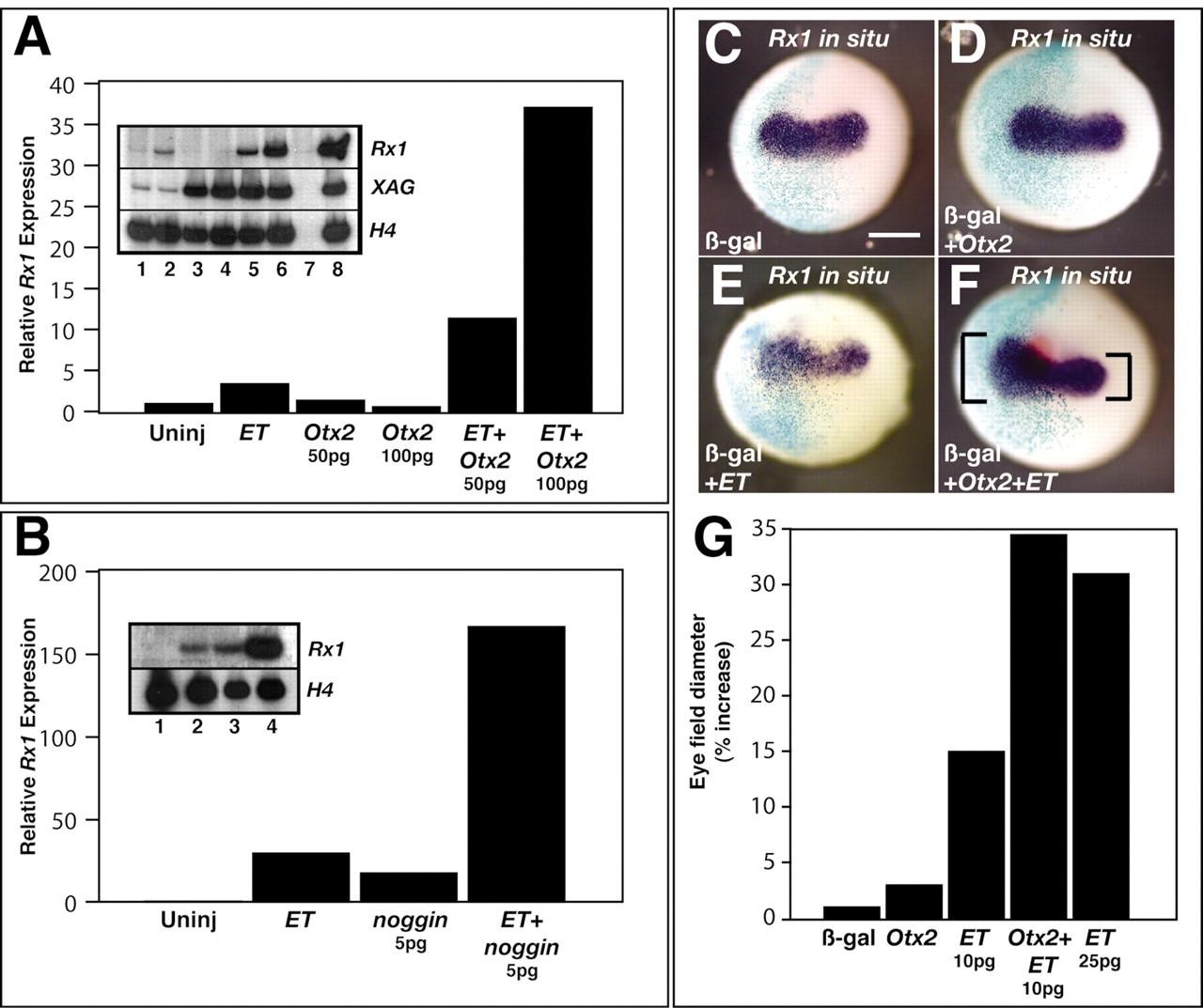
Fig. 6. Otx2 and noggin potentiate the induction of Rx1 by ET. (A,B) RT-PCR was used to detect changes in Rx1 and XAG expression in ectodermal explants from Xenopus embryos injected with noggin, Otx2 and ET. ET (100 pg) was injected alone, with 50 or 100 pg of Otx2 (A), or 5 pg noggin (B). (A) Lane 1, uninjected; lane 2, ET (100 pg); lane 3, Otx2 (50 pg); lane 4, Otx2 (100 pg); lane 5, ET (100 pg) + Otx2 (50 pg); lane 6, ET (100 pg) + Otx2 (100 pg); lane 7, embryo, no reverse transcription; lane 8, embryo, XAG induction was used as a positive control for Otx2 activity. (B) Lane 1, uninjected; lane 2, ET (100 pg); lane 3, noggin (5 pg); lane 4, ET (100 pg) + noggin (5 pg). (C-G) Rx1 expression was normalised to Histone H4 then set relative to uninjected controls. Otx2 potentiates the ET induced expansion of Rx1 expression in the anterior neural plate. Whole-mount in situ hybridisation was used to detect Rx1 expression at stage 13 in embryos injected with βgal alone (C), or in combination with 25 pg Otx2 (D), 10 pg ET (E) or both Otx2 and ET (F). (G) The rostrocaudal diameter of the Rx1 expression domain on the injected side (βgal-positive) was measured and compared with the uninjected (βgal-negative) side of the embryo (see F for an example). Image published in: Zuber ME et al. (2003) Copyright © 2003. Image reproduced with permission of the publisher and the copyright holder. This is an Open Access article distributed under the terms of the Creative Commons Attribution License.
Image source: Published Larger Image Printer Friendly View |