XB-IMG-24040
Xenbase Image ID: 24040
|
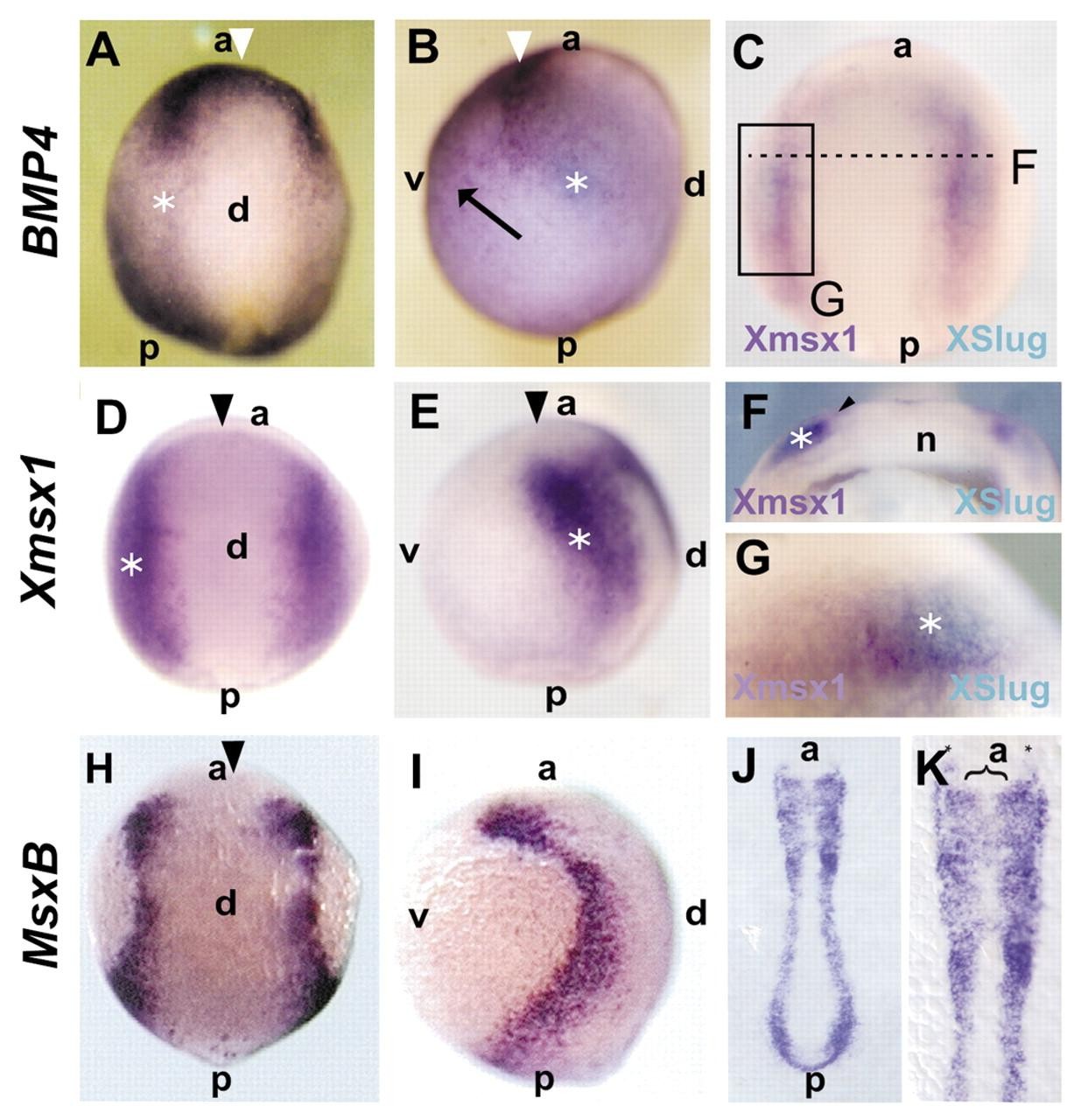
Fig. 1. Msx genes are expressed in the neural crest region of Xenopus and zebrafish embryos. (A-G) Xenopus embryos. Stage 13. (H-K) Zebrafish expression. Bud and five-somites stage. Anterior is upwards. (A,B) Bmp4 expression. (C-G) msx1 expression. (H-K) msxb expression. Arrowhead indicates anterior neural fold; asterisk: indicates prospective neural crest. (A) Dorsal view, showing strong expression in the anterior neural fold. (B) Lateral view, showing strong expression in the anterior neural fold, intermediate in the prospective neural crest and weaker in the ventral ectoderm. Arrow in B indicates expression in the ventral side. (C) Dorsal view of a double in situ hybridization for msx1 (purple) and XSlug (blue) of a mid/late gastrula stage embryo. Note the overlapping in the expression of both genes in the prospective neural crest region (square) in the whole embryo (C), in the sectioned embryo (F) and in a higher magnification (G). (D) Dorsal view showing the strong expression of msx1 in the neural folds. (E) Lateral view showing expression in the neural folds. (H) Dorsal view showing expression of msxb in the neural plate border. (I) Lateral view showing expression in the neural plate border. (J) Flat mount showing expression in the prospective neural crest region. (K) Higher magnification of J. Image published in: Tribulo C et al. (2003) Copyright © 2003. Image reproduced with permission of the Publisher and the copyright holder. This is an Open Access article distributed under the terms of the Creative Commons Attribution License.
Image source: Published Larger Image Printer Friendly View |