XB-IMG-26221
Xenbase Image ID: 26221
|
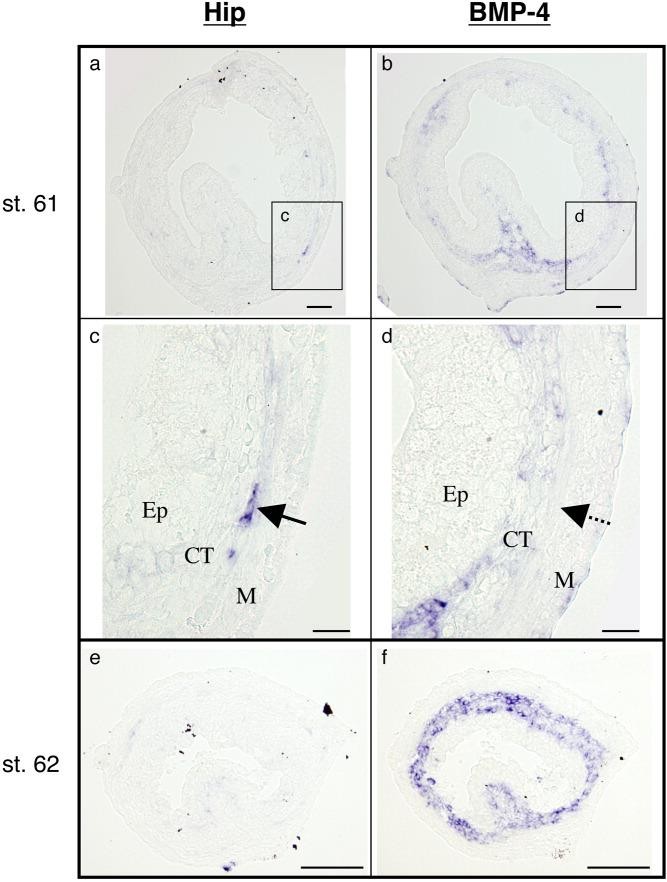
Figure 3. Comparison of hedgehog interacting protein (Hip) and bone morphogenetic protein (BMP) -4 localization. Cross-sections of the intestine at stage 61 (a-d) and stage 62 (e,f) were hybridized with antisense Hip (a,c,e) and BMP-4 (b,d,f) probes. To compare the localization of Hip with that of BMP-4, serial sections were used. Dark blue deposits indicate the sites of probe binding. Light or dark brown pigments are melanin. c,d: Higher magnification of a boxed area in a and b, respectively. c,d: The signals for BMP-4 mRNA (d, dashed arrow) are hardly detectable where Hip is strongly expressed (c, solid arrow). BMP-4 is highly expressed at stage 62 (f) when the expression of Hip decreases (e) and that of Shh peaks (Fig. 2c). Ep, epithelium; CT, connective tissue; M, muscle layer. Scale bars = 100 mu m in a,b,e,f, 20 mu m in c,d. Image published in: Hasebe T et al. (2008) Copyright © 2008. Image reproduced with permission of the Publisher, John Wiley & Sons.
Image source: Published Larger Image Printer Friendly View |