XB-IMG-2966
Xenbase Image ID: 2966
|
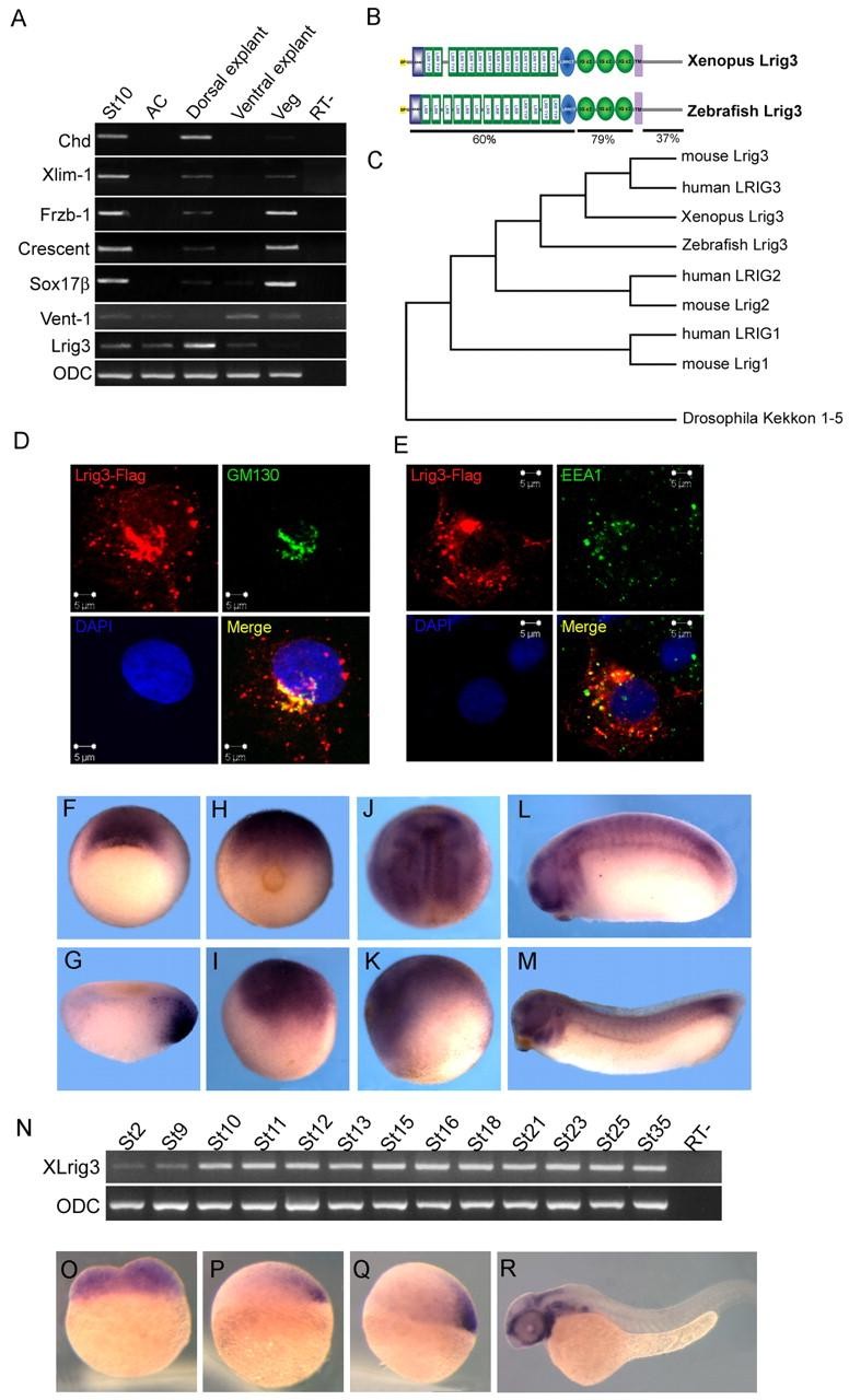
Fig. 1. Characterization of the Lrig3 gene. (A) Regional expression of Lrig3 in stage 10 embryos. Lrig3 was expressed strongly in dorsal explants, but barely detected in other regions. The indicated genes were assayed to test quality of dissection. St10, stage 10 embryo; AC, animal cap; Veg, vegetal explant; RT-, control without reverse transcriptase. (B) Schematic drawing of the protein structure of Xenopus and zebrafish Lrig3. SP (yellow), signal peptide; LRRNT (blue box), leucine-rich repeat N-terminal domain; LRR TYP, leucine-rich repeats, typical; LRR, leucine-rich repeats; LRRCT (blue oval), leucine rich repeat C-terminal domain; IG C2 (green oval), immunoglobulin C-2 Type; TM (purple), transmembrane domain. Sequence identity between zebrafish and Xenopus Lrig3 is indicated. (C) Dendrogram of the Lrig3 family including Kekkon of Drosophila. (D,E) Subcellular distribution of Lrig3 after transfection into COS7 cells. (D) Transfected Lrig3-Flag (red) co-localized with the cis Golgi apparatus marker GM130 (green); the nucleus was stained with DAPI (blue). (E) A small proportion of Lrig3-Flag co-localized with the early endosome marker EEA1 (green). (F-M) Expression pattern of Lrig3 in Xenopus. Vegetal view at stage 10, expression in the organizer (F); stage 10 section (G). (H,I) Stage 12 (H, posterior view; I, lateral view). (J,K) Stage 15 (J, dorsal view; K, lateral view). Tailbud (stage 24, L) and tadpole (stage 32, M); expression is seen in brain, eye, somites and branchial arches. (N) Temporal expression of Lrig3 during Xenopus development. (O-R) Expression of lrig3 in zebrafish. Transcripts are present maternally (O), become localized in the forming organizer at 30% epibody (P) and subsequently in the shield (Q), and were found in brain, eye and branchial arches at 24 hours (R). (P-R) Lateral views. Image published in: Zhao H et al. (2008) Copyright © 2008. Image reproduced with permission of the Publisher and the copyright holder. This is an Open Access article distributed under the terms of the Creative Commons Attribution License.
Image source: Published Larger Image Printer Friendly View |