XB-IMG-30668
Xenbase Image ID: 30668
|
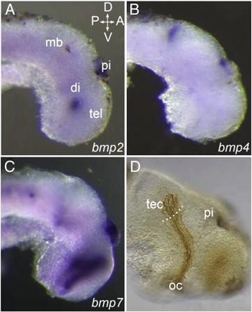
Fig. 8. BMP ligands are expressed near the developing optic tract. (AâC) Stage 35/36 wholemount brains processed for in situ hybridization using antisense probes to bmp2 (A), bmp4 (B), and bmp7 (C). All three mRNAs are expressed near or within the pineal gland. bmp2 is also expressed in a spot in the mid-diencephalon (A), and bmp4 in a stripe at the back of the tectum (B). bmp7 is expressed in a large area of the ventral diencephalon and telencephalon (C). (D) Wholemount stage 40 Xenopus brain with RGC axons labelled brown following GFP electroporation and anti-GFP immunochemistry. This shows the optic pathway for comparison to the bmp expression domains. The dotted white line is at the approximate tectal border. A, anterior; D, dorsal; di, diencephalon, mb, midbrain; oc, optic chiasm; pi, pineal gland; P, posterior; tec, tectum; tel, telencephalon; V, ventral. Image published in: Hocking JC et al. (2009) Copyright © 2009. Image reproduced with permission of the Publisher, Elsevier B. V.
Image source: Published Larger Image Printer Friendly View |