XB-IMG-41485
Xenbase Image ID: 41485
|
|||||||||||||||||||||||||||||||||||
|
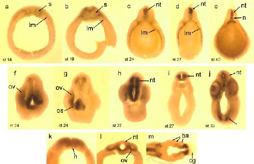
Fig. 3. Vibratome sections of embryos stained by whole mount in situ hybridization with FGFR3 probe. (a and b) Sections through the level of the future hindbrain, at stage 14 (a) and 19 (b). (câe) Sections through the trunk at stages 24 (c), 27 (d) and 40 (e). (f and g) Adjacent sections through the head of a stage 24 embryo, showing staining in the medial part of the optic vesicle and optic stalk. (h and i) Sections through the head of a stage 27 embryo, showing strong staining throughout the neural tube. The section in (i) is between the eyes and otic vesicles. Note the absence of staining in the roof plate of the neural tube. (j) Section through the head of a stage 33 embryo, showing staining in the ventricular surface of the neural tube, the medial eye, lens, and in the ventral part of the head. Arrow indicates cells in the ventral head, near the cement gland. (kâm) Frontal sections through embryos at stages 19 (k), 24 (l), and 33 (m). Anterior is up in (k) and (l); anterior is to the right in (m). The section in (k) cuts through the neural tube as it curves around the anterior of the embryo (cf. Fig. 2e), the section in l cuts through the optic vesicles, and the section in m is ventral to the eyes, through the cement gland. abbreviations: ba, branchial arches; cg, cement gland; lm, lateral mesoderm; n, notochord; nt, neural tube; os, optic stalk; ov, optic vesicle; s, somite. Image published in: Pope AP et al. (2010) Copyright © 2010. Image reproduced with permission of the Publisher, Elsevier B. V.
Image source: Published Larger Image Printer Friendly View |