XB-IMG-42246
Xenbase Image ID: 42246
|
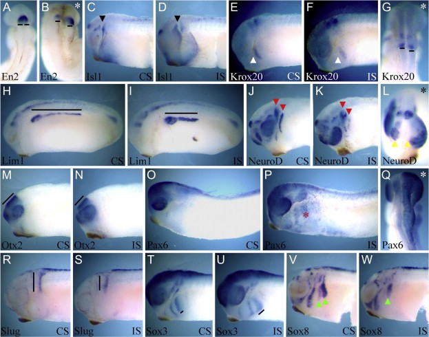
Fig. 3. Overexpression of XtSulf2 affects the expression of a number of genes expressed in anterior neural tissue and the cranial neural crest. One blastomere of a two-cell Xenopus tropicalis embryo was injected with 0.5Â ng of XtSulf2 mRNA, embryos were allowed to develop to NF stage 28 and fixed for in situ hybridization using En2 (AâB), Isl1 (CâD), Krox20 (EâG), Lim1 (HâI), NeuroD (JâL), Otx2 (MâN), Pax6 (OâQ), Slug (RâS), Sox3 (TâU), and Sox8 (VâW). En2 expression is shifted posteriorly on the injected side (AâB). Isl1 expression in the profundal and trigeminal placodes shifts posteriorly (black arrowheads) as does the expression in the heart (CâD). Krox20 expression in the migrating neural crest shifts posteriorly (EâF white arrowheads) as does the expression in r5 (G black lines). Lim1 expression in the pronephros is restricted anteriorly (HâI black lines). NeuroD expression in the profundal and trigeminal placodes is disrupted (JâK red arrowheads), and expression in the olfactory placodes fuses with expression in the eye placodes (L yellow arrowheads). Otx2 expression is restricted in the midbrain (MâN black lines). Pax6 expression is ectopically expressed in the posterior and dorsal head ectoderm (OâPâQ red asterisk). Slug expression at the leading edge of neural crest migration remains closer to the neural tube than in controls (RâS black lines). Sox3 expression is decreased in neural crest but expands laterally (TâU black lines). Sox8 expression in the anterior and posterior branchial arches is decreased and the leading edge of the migrating neural crest remains closer to the neural tube than in controls (VâW green arrowhead). (AâB, G, Q) are dorsal views of embryos, anterior at the top. (CâF, HâK, MâP, RâW) are lateral views of embryos, anterior to the left. (L) is an anterior view of embryo in (JâK), dorsal at the top. Asterisks indicate injected side. CS control side, IS injected side. Image published in: Guiral EC et al. (2010) Copyright © 2010. Image reproduced with permission of the Publisher, Elsevier B. V.
Image source: Published Larger Image Printer Friendly View |