XB-IMG-42571
Xenbase Image ID: 42571
|
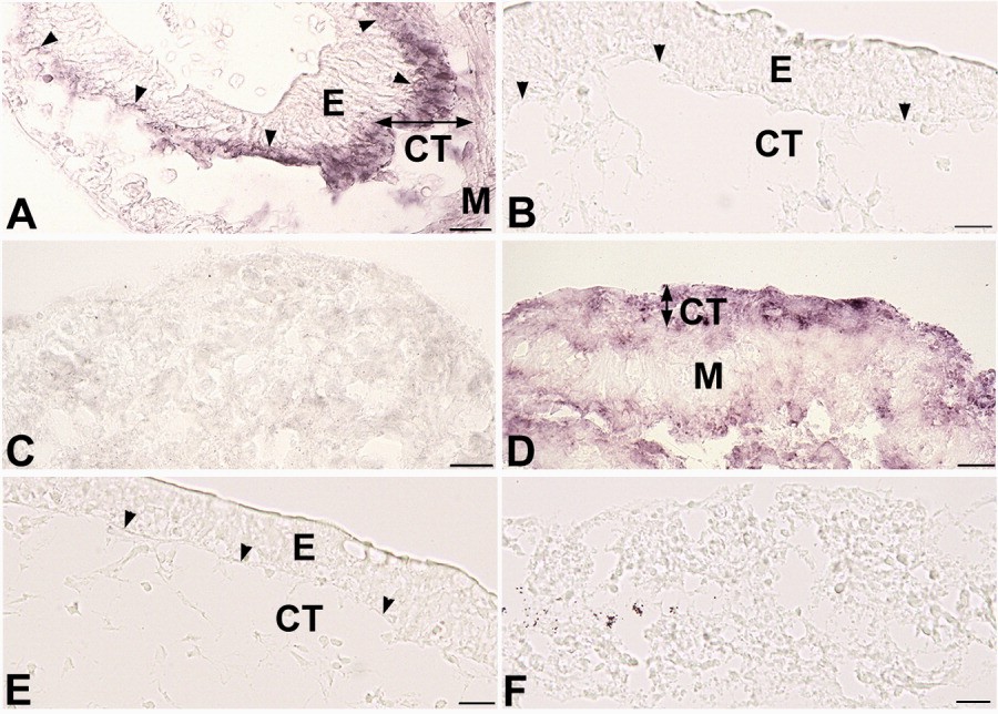
Figure 1. In vitro expression of bone morphogenetic protein-4 (BMP-4) mRNA in Xenopus laevis intestines cultured for 5 days and analyzed by in situ hybridization. A,B: Intact intestines. A high level of BMP-4 mRNA is localized in the connective tissue (CT) just beneath the epithelium (E) in the presence of thyroid hormone (TH, A) but not in its absence (B). Small arrowheads show the boundary between the epithelium and the connective tissue. C,D: Epithelium-free intestines cultured in the presence of TH. TH up-regulation of BMP-4 expression does not occur in the absence of Shh (C). When Shh protein (500 ng/ml) is added to the medium (D), BMP-4 mRNA becomes detectable in the connective tissue but does not in muscles (M). E,F: Intact (E) and epithelium-free (F) intestines cultured in the absence of TH. Even though Shh is added to the medium, BMP-4 mRNA remains undetectable in any tissue. Scale bars = 20 mu m. Image published in: Ishizuya-Oka A et al. (2006) Copyright © 2006. Image reproduced with permission of the Publisher, John Wiley & Sons.
Image source: Published Larger Image Printer Friendly View |