XB-IMG-43976
Xenbase Image ID: 43976
|
||||||||||
|
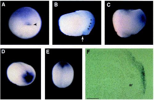
Fig. 3. Xfz8 mRNA is localized in dorsal and anterior regions of early embryos. Whole mount in situ hybridization was carried out with antisense Xfz8 RNA probe for embryos of different developmental stages. (A) Stage 10.5 gastrula, dorsovegetal view. Staining in the dorsal lip is marked by an arrowhead. (B) Midgastrula embryo was cut in half along mid-sagittal plane after in situ hybridization to show neuroectoderm staining. Arrowheads point to the interface between neuroectoderm and underlying mesoderm; white arrow indicates dorsal blastopore. (C) Stage 11.5 gastrula, dorsal is on the right. (D,E) Stage 19 neurula, anterolateral (D), and dorsal (E) views. Most anterior staining is apparent. (F) Stage 19 neurula, sagittal section, anterior is to the right. ar, archenteron. Scale bar, 200 μm. Image published in: Itoh K et al. (1998) Copyright © 1998. Image reproduced with permission of the Publisher, Elsevier B. V.
Image source: Published Larger Image Printer Friendly View |