XB-IMG-47131
Xenbase Image ID: 47131
|
Fig. 1. Zygotic expression of XtSulf1 analysed by whole mount in situ hybridisation. (A) Stage 13 embryo shows XtSulf1 expression in the posterior paraxial mesoderm (dorsal view;
anterior to the left). (B) Stage 20 embryo shows XtSulf1 expression in the pre-segmented posteriormesoderm, the somites, and the ventral part of the hind brain and the floorplate of
the neural tube (lateral view; anterior to the left). (C) Stage 26 embryo shows expression in the posteriormesoderm, the somites, the hindbrain, the floorplate of the neural tube, the
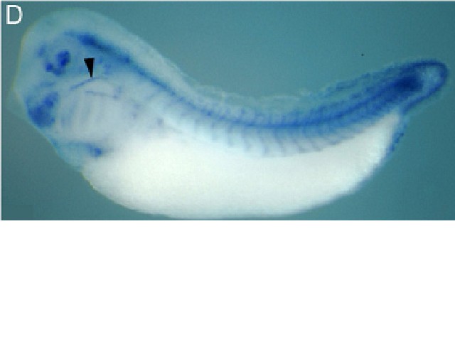
retina of the eye, some neural crest (arrowhead), and the pronephros (arrow). (D) At later tailbud stages, XtSulf1 is expressed in the mesoderm of the tailbud, the somites, the
hindbrain, the neural crest (arrowhead), the floorplate of the neural tube, the fin, the pericardium, the lens, the ciliary marginal zone (CMZ) of the retina, and in the first branchial
arch. (E) Vibratome section through D showing expression of XtSulf1 in the ventricular part of the midbrain, the lens (arrowhead) and CMZ of the eye (arrow). (F) Vibratome section
through C showing the expression in the pronephros (arrowhead) and the floorplate (arrow). (G) Vibratome section of D showing the expression of XtSulf1 in the somites and
floorplate. This image is extracted from figure published in: Freeman SD et al. (2008), Image published in: Freeman SD et al. (2008) Copyright © 2008. Image reproduced with permission of the Publisher, Elsevier B. V.
Image source: Published Larger Image Printer Friendly View |