XB-IMG-48085
Xenbase Image ID: 48085
|
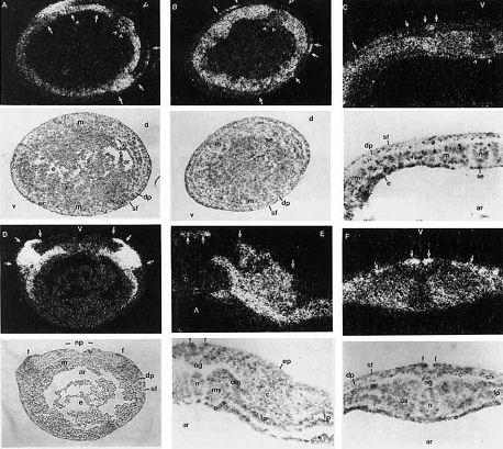
Fig, 4. Xsna expression during Xenopus neurulation. Distribution of
Xsna mRNA in neurulae shown by in situ hybridisation under dark field
illumination, with corresponding bright field views below. Single large
arrowheads, dorsal midline; small arrowheads, Xsna-expressing supra-
blastoporal endoderm; arrows, ectodermal Xsna expression. Scale bar =
400 pn in A,B,D; 200 pm in C,E,F. A: Stage 12. Anterior transverse
section. Ring of mesodermal mantle (m) and sub-notochordal endoderm
(se, small arrowheads) are labelled. Cross sections of stripes of Xsna
expression (arrows) in deep (dp) and superficial (sf) ectoderm shown,
symmetrical about midline (large arrowhead). 8: Stage 12. Transverse
section near blastopore of embryo in (A). Gap in labelling of mesodermal
mantle (m) on dorsal midline (large arrowhead) reveals prospective no-
tochord (n). Stripe of Xsna expression in deep ectoderm (dp, arrows-
seen only on right, as left-ventral shows un-involuted mesoderm) is ven-
tral (v) and separate from superficial ectoderm (sf) expression (arrows).
C: Stage 13. Prospective trunk transverse section. Stripes of Xsna ex-
pression (arrows) in superficial (sf) and deep (dp) ectoderm are nearer
dorsal midline (large arrowhead). All mesoderm (m) except notochord (n)
is labelled. Archenteron (ar) roof except sub-notochordal endoderm (se,
small arrowheads) is unlabelled. D: Stage 14. Prospective head trans-
verse section. Stripes of ectodermal Xsna expression (arrows) are in
neural folds (f) on either side of neural plate (np). E: Stage 18. Transverse
section at level of head somites. Like notochord (n), rotated myotome
(my) has become unlabelled, leaving dorso-lateral and ventro-medial
somite compartments, probably dermatome (dm) and sclerotome (s), and
both layers of lateral plate mesoderm (Ip) labelled. Superficial ectoderm
on tips of neural folds (f), almost touching above neural groove (ng), and
lateral aggregate of pre-migratory cephalic neural crest (c) are also la-
belled. F: Stage 18. Mid-trunk transverse section of embryo in (E). Pos-
terior paraxial mesoderm either side of unlabelled notochord (n) is un-
segmented (us) and, like lateral plate mesoderm (Ip), still contains Xsna
transcripts. Xsna-expressing ectodermal cells occupy same positions as
in the head (see E; f, c), but deep layer (dp) expression zone is very thin.
ar = archenteron; c = neural crest; d = dorsal; dm = dermatome; dp =
deep ectoderm; e = endoderm; ec = ectoderm; ep = epidermis; f =
neural fold; Ip = lateral plate mesoderm; m = mesoderm; my = myo-
tome; n = notochord; ng = neural groove; np = neural plate; s =
sclerotome; se = sub-notochordal endoderm; sf = superficial ectoderm;
us = unsegmented paraxial mesoderm; v = ventral. Image published in: Essex LJ et al. (1993) Copyright © 1993. Image reproduced with permission of the Publisher, John Wiley & Sons.
Image source: Published Larger Image Printer Friendly View |