XB-IMG-48753
Xenbase Image ID: 48753
|
|||||||||||||||||||||||||
|
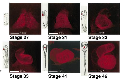
FIG. 1. Cardiac muscle development in Xenopus. Confocal images of the heart regions of six stages of embryos (27-46) are presented (ventral views), each adjacent to a diagrammatic representation of an embryo (Nieuwkoop and Faber, 1967) at the corresponding stage. Prior to microscopy, stage 27-33 embryos were immunolabeled with anti-tropomyosin (CH1) and stages 356 with anti-cardiac troponin T (CT3). The secondary antibody used for all stages was conjugated to Cy5. Each confocal image represents a digitally colored compilation of 30-53 optical sections taken (in gray scale) 5 um apart. All confocal images were taken at the same magnification and are presented with the anterior of the embryo at the top. Scale bar, 100 um Image published in: Kolker SJ et al. (2000) Copyright © 2000. Image reproduced with permission of the Publisher, Elsevier B. V.
Image source: Published Larger Image Printer Friendly View |