XB-IMG-48872
Xenbase Image ID: 48872
|
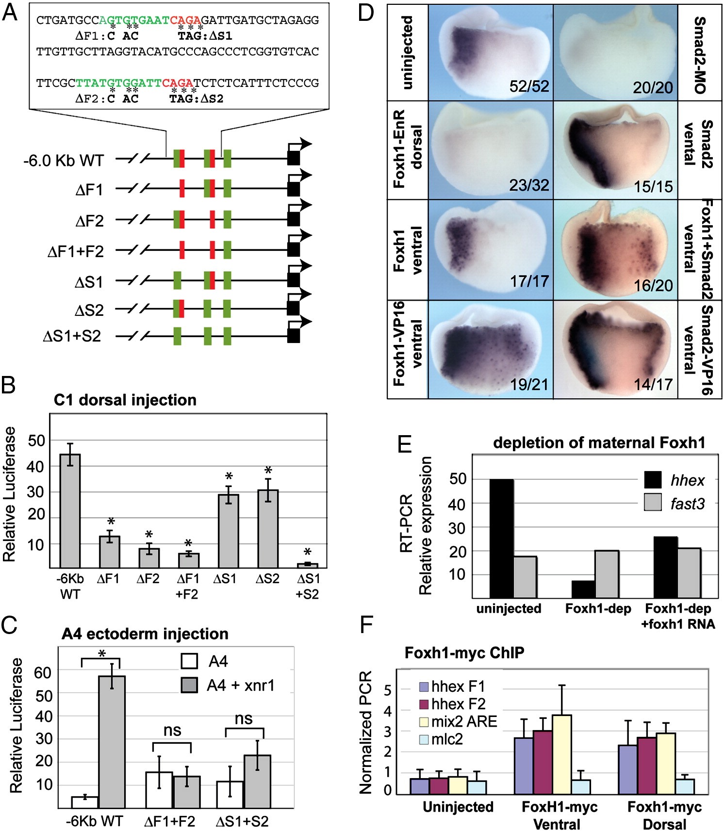
Fig. 5. Nodal-signaling directly activates hhex transcription through Foxh1/Smad2 binding in the proximal â0.44 Kb promoter. (A) Schematic and sequence of the NRE indicating the putative Foxh1 (green) and Smad (red) DNA-binding sites and corresponding âF and âS mutations. (BâC) Relative luciferase activity in gastrulae injected with the indicated reporter constructs into (B) the C1 dorsal mesendoderm or (C) the A4 ectoderm along with or without xnr1 RNA (50 pg). Histograms show the average normalized luciferase activity and standard deviation from a single injection experiment performed in biological triplicate. A representative from 3 independent experiments is shown. *p < 0.01 in Student T-test in (B) compared to the wt reporter or in (C) compared to injection of the same reporter alone; ns = no statistical difference. (D) Foxh1 and Smad2 regulate endogenous hhex expression. Hhex in situ of gastrulae injected into either dorsal or ventral cells with: foxh1-EnR RNA (500 pg), foxh1-VP16 RNA (200 pg), Smad2a/b -MOs (20 ng each), Smad2-VP16 RNA (200 pg) foxh1 RNA (250 pg), smad2 RNA (250 pg), or foxh1 + smad2 RNAs (125 pg each). (E) Normalized QRT-PCR of hhex and fast3 mRNA levels in gastrulae depleted of maternal Foxh1 and rescued by co-injection of foxH1 RNA (100 pg). (F) Normalized QPCR analysis of chromatin immunoprecipitated (in triplicate) from gastrulae injected dorsally or ventrally with Myc-Foxh1 RNA (50 pg). Primers amplified genomic DNA fragments containing the F1 or F2 Foxh1-sited in the hhex NRE, the mix2 ARE as a positive control and mlc2 promoter as a negative control. Image published in: Rankin SA et al. (2011) Copyright © 2011. Image reproduced with permission of the Publisher, Elsevier B. V.
Image source: Published Larger Image Printer Friendly View |