XB-IMG-49452
Xenbase Image ID: 49452
|
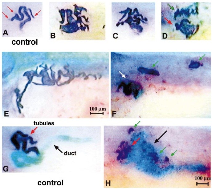
FIG. 3. Ectopic expression of Pax-8 plus Xlim-1 leads to the development of abnormally large pronephroi and ectopic pronephroi. All samples were stained with antibody 3G8. A reproduced at same magnification, as are G and H (100-mmscale bars are in E and H). Anterior
is to the left, dorsal is up. (A) Normal stage 36 pronephric tubules. Red arrows indicate the three normal dorsal branches. Faintly stained nephrostomes can be seen extending from the two left branches. (B, G) Enlarged pronephroi in XPax-8 plus Xlim-1- (1:1 ratio) injected embryos. In B the tubules to the left are slightly distended, probably due to osmotic pressure, and this thickness probably represents distortion rather than enlargement. However, the right side of this same pronephros contains many additional tubule branches that are all
of normal thickness. In C, all tubules are of normal width. In D the pronephros (red arrow) is of only slightly greater than normal size, but it is adjacent to an ectopic pronephros (green arrow) which is almost as large. In F, two additional ectopic pronephroi (green arrows) are obvious, dorsal and posterior to the normal position of the organ (white arrow). (G) Control stage 39 embryo stained for pronephric tubules
using 3G8 and a dark blue substrate and pronephric duct using 4A6 and a light blue substrate. Note that G and H are at a later stage of development than A, and the scale is also slightly different. (H) XPax-8 plus Xlim-1-injected embryo, stage 39, stained as in G. Note the enlarged region of nonmigratory duct staining (light blue) in the vicinity of the pronephric tubules and also the presence of small ectopic pronephric tubules (dark blue stain, green arrows). Anterior is to the left, and dorsal is up in all panels. Image published in: Carroll TJ and Vize PD (1999) Copyright © 1999. Image reproduced with permission of the Publisher, Elsevier B. V. Larger Image Printer Friendly View |