XB-IMG-49648
Xenbase Image ID: 49648

|
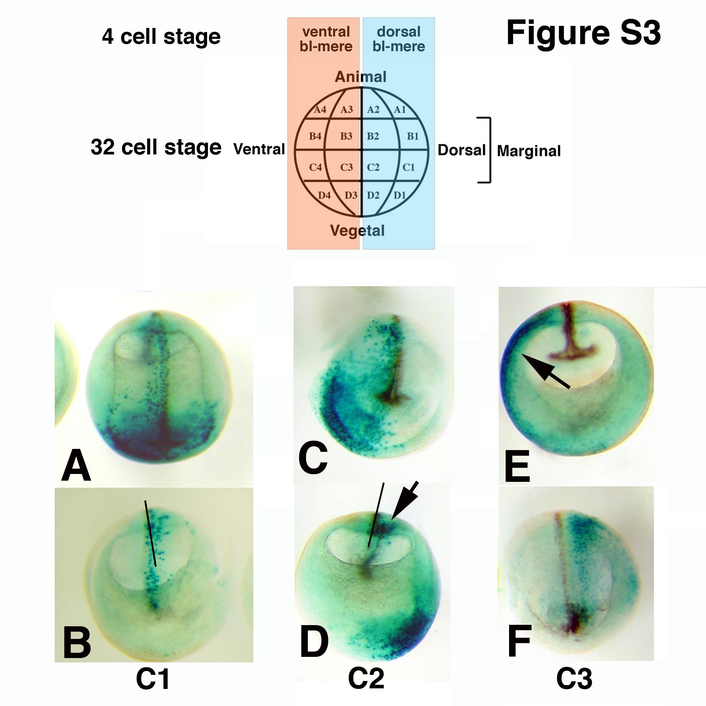
Supplementary Fig. S3. Cell fate of tier C blastomeres from 32 cell stage embryos, at stage 18. 32 cell stage embryos (upper panel, nomenclature of Nakamura, descendents of the dorsal blastomere from 4 cell stage in light blue, and of the ventral blastomere in pink) were injected in blastomeres C1, C2 (dorsal descendents) and C3 (ventral descendent) with 2 ng LacZ RNA, collected at stage 18, and stained with X-Gal (blue dots). All embryos are cleared. A, B. C1 injection. A: dorsal view. B: sectioned posterior half, internal view. Most of the tracer migrates to the anterior pole, the rest is mostly axial, with some contribution to posterior paraxial mesoderm. C, D. C2 injection. C: anterior half, external view. D: posterior half, internal view. Arrow in D indicates tracer in ventral paraxial mesoderm. E, F. C3 injection. E: anterior half, internal view. Arrow indicates lateral plate mesoderm. F: posterior half, external view. Tracer is lateral to the posterior paraxial mesoderm. Bars in B and D indicate the midline. Image published in: Vonica A and Brivanlou AH (2007) Copyright © 2007. Image reproduced with permission of the Publisher, Elsevier B. V. Larger Image Printer Friendly View |
