XB-IMG-74200
Xenbase Image ID: 74200
|
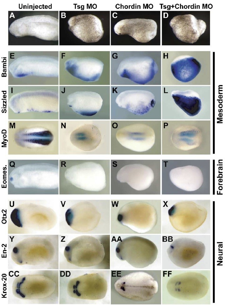
Fig. 5. Depletion of chordin and tsg reduces dorsal tissues and results in a cooperative expansion of ventral tissues. Embryos were injected at the two-cell stage with tsg MO (B, F, J, N, R, V, Z, DD), chordin MO (C, G, K, O, S, W, AA, EE), or both MOs (D, H, L, P, T, X, BB, FF) and compared to uninjected sibling control embryos (A, E, I, M, Q, U, Y, CC). Embryos were assayed for expression of the ventral markers BAMBI (E H) and sizzled (I L) and the somite marker myoD (M P). Embryos were also assayed for expression of several regional specific markers of the neural plate: eomesodermin (Q), otx2 (U), en-2 (YB) and krox-20 (CC FF). All embryos are between stages 22 25. Embryos stained for expression of myoD (M P) and krox -20 (CC FF) are viewed dorsally with anterior to the left, all others are viewed laterally with anterior to the left. Image published in: Wills A et al. (2006) Copyright © 2006. Image reproduced with permission of the Publisher, Elsevier B. V.
Image source: Published Larger Image Printer Friendly View |