XB-IMG-74404
Xenbase Image ID: 74404
|
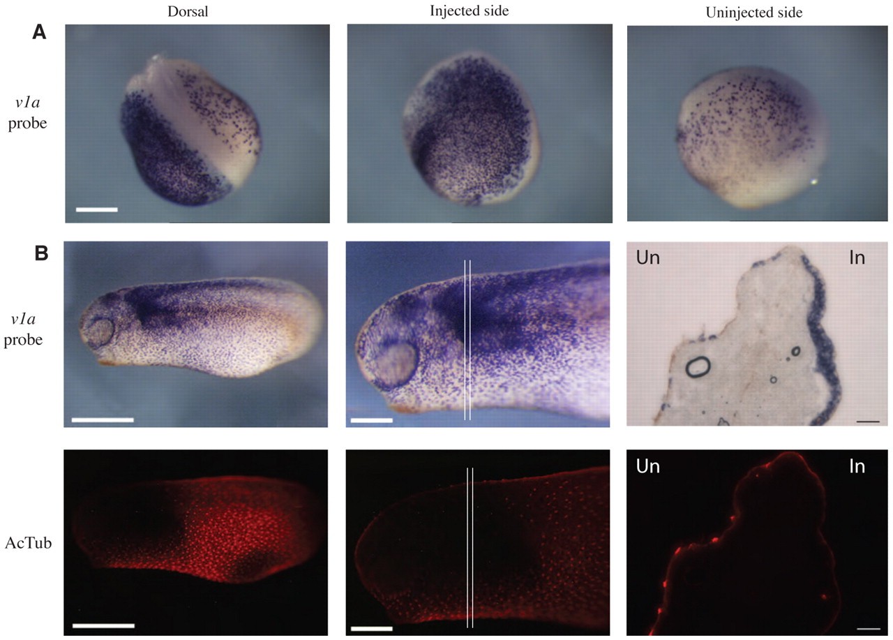
Fig. 5. Misexpression of foxi1e causes an increase in ionocytes at the expense of ciliated cells. (A) Embryos injected with foxi1e-HA RNA (500 pg) and lacZ (marks injected side; blue) were hybridised with the v1a probe at neurula stage. Whole-mount embryo is shown in the views indicated. Scale bar: 250 μm. (B) The injected side of an embryo shown at low magnification (left), high magnification (middle) and in cross-section (right). Upper and lower panels represent the same field of imaging, with upper panels showing in situ hybridisation with the v1a probe (purple) and lower panels showing antibody staining for the ciliated cell marker acetylated α-tubulin. Parallel lines in the high-magnification images show region that was sectioned. Sectioned images show an abundance of v1a ionocytes on the injected side (In) compared with the uninjected side (Un). Ciliated cells are absent in regions overexpressing v1a and this is emphasised by comparison of the injected side with the uninjected side in cross-sections. Scale bars: 500 μm (low magnification); 250 μm (high magnification); 100 μm (sections). Image published in: Dubaissi E and Papalopulu N (2011) © 2011. Creative Commons Attribution-NonCommercial-ShareAlike license
Image source: Published Larger Image Printer Friendly View |