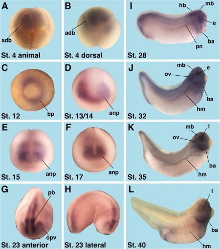
Figure 2. Analysis of the spatial expression pattern of X. laevis IRS-1. Whole-mount in situ hybridizations at the indicated stages. adb, animal dorsal Bugner V et al. (2011) Gene Synonyms Species Stage(s) Tissue irs1.L irs-1, LOC108718134 X. laevis Throughout NF stage 12 irs1.L irs-1, LOC108718134 X. laevis Throughout NF stage 13 pre-chordal neural plate irs1.L irs-1, LOC108718134 X. laevis Throughout NF stage 15 pre-chordal neural plate irs1.L irs-1, LOC108718134 X. laevis Throughout NF stage 17 pre-chordal neural plate irs1.L irs-1, LOC108718134 X. laevis Throughout NF stage 23 optic vesicle irs1.L irs-1, LOC108718134 X. laevis Throughout NF stage 28 eye irs1.L irs-1, LOC108718134 X. laevis Throughout NF stage 32 eye irs1.L irs-1, LOC108718134 X. laevis Throughout NF stage 35 and 36 eye irs1.L irs-1, LOC108718134 X. laevis Throughout NF stage 40 branchial arch irs1.L irs-1, LOC108718134 X. laevis Throughout NF stage 4 (8-cell) animal blastomere
Image source: PublishedLarger Image Printer Friendly View