XB-IMG-75075
Xenbase Image ID: 75075
|
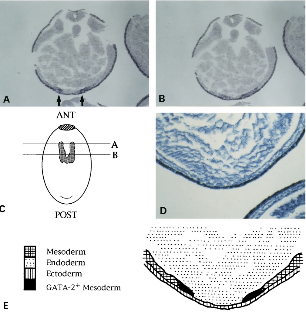
Fig. 3. GATA-2 is localised in presumptive haematopoietic cells before globin expression is established. Late neural tube (stage 21) non-albino embryos were hybridised with an antisense GATA-2 probe. (A,B,D,E) The orientation of the embryos is ventral side down. (A) Transverse section through the anterior end of the presumptive blood islands showing ventral position and two discrete expression domains (arrows). (B) Section taken just posterior to (A) showing the two discrete expression domains closer together. (C) Schematic diagram of the ventral aspect of a stage 21 embryo showing the mesodermal expression of GATA-2 (stippled area) reconstructed from examination of serial sections. Note the of the future blood islands. For orientation, the cement gland (hatched area, ANT) and blastopore (curve, POST) are shown.
(D) Section from the same region as in (A), at higher power, stained with brilliant cresyl blue for histological detail. (E) A schematic illustration of the section shown in (D) to indicate the three primary germ layers. Note that in this position along the anteroposterior axis of the embryo, the mesoderm does not extend to the ventral midline. Areas of GATA-2 expression (shown in black) are restricted to the medial edges of the mesoderm in this region. Image published in: Walmsley ME et al. (1994) Copyright © 1994. Image reproduced with permission of the publisher and the copyright holder. This is an Open Access article distributed under the terms of the Creative Commons Attribution License. Larger Image Printer Friendly View |